


INTRODUCTION
The image analysis method has been developed for objective evaluation of meat quality (Tan, 2004). It can also be applied for the evaluation of meat marbling (Albrecht et al., 1996; Gerrard et al., 1996; Kuchida et al., 2000; Yang et al., 2006). Marbling in beef is an important factor that influences taste, juiciness, tenderness and flavor (Okitani, 2002; Platter et al., 2005). Wagyu beef, in particular, served in the Japanese restaurants in Asia, China, Europe and USA often possesses abundant marbling, the market rate thus sometimes exceeds $20/kg of carcass or $100/kg of retail loin, depending mainly on marbling. Although graders are professionally trained, subjective evaluation often lead to inconsistencies and variations (Cross et al., 1983).
The image analysis method for marbling has been developed but not yet been applied in meat grading for highly marbled beef in Asia, because it is labor consuming for capture of image and manual operation of correction and needs a special apparatus. Original image analysis method is extremely rapid, and its automation has been advanced. Image analysis for beef marbling is used in North America (personal comm). However, when the muscle outline is auto-traced for evaluation of high marbling, troublesome manual operation is often needed for distinguish between meat and fat, especially for highly marbled meat such as that of Wagyu beef.
Therefore, we designed a spot method that measures a small area of a constant part without the need for manual correction. Moreover, this method has the merit of evaluating the degree of marbling even when only partial images are obtained from a narrowly cut section to prevent abnormal diffusion of prions or when the evaluation of other portions such as the hip is required for determining the accurate commercial value. Further, we attempted to compare the overall trace method with the spot method in evaluating the marbling of each beef muscle.
MATERIALS AND METHODS
Sample preparation
Samples were obtained from carcasses of Wagyu (Japanese Black) cattle. Its beef is characterized by a unique ability to deposit large amounts of intramuscular fat (Zembayashi et al., 1988; Cameron et al., 1994). The materials included 82 cross sections at the 6 to 7th rib of carcasses, which is the Japanese standard location for measuring marbling.
Photographs of the loin cuts including longissimus thoracis, trapezius, semispinalis dorsi and semispinalis capitis muscles were taken with a scale using a digital camera (C-3100, Olympus Co., Tokyo, Japan) on the carcasses at meat markets. The flash unit in the digital camera was not used because the light reflectance of moisture on meat surface lead to error.
Image analysis
The photographic images were imported to a personal computer loaded with the image analysis software (Image-Pro PLUS Ver. 4.0, Media Cybernetics, Inc., MD, USA). The images were calibrated with the scale in the photograph according to the software requirements. To eliminate subjective operator-to-operator differences, the measurements were performed by only 1 experienced operator.
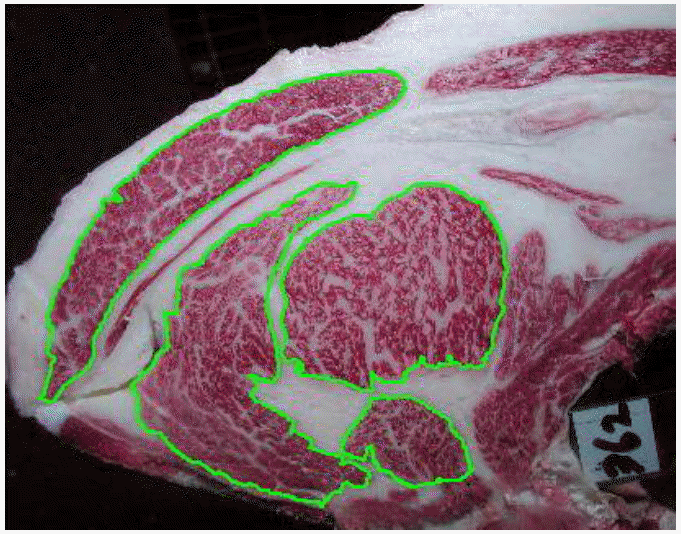
In the overall trace method, the surrounding edges of the longissimus thoracis, trapezius, semispinalis dorsi and semispinalis capitis muscles were automatically traced by auto trace operation in the image analysis software (Figure 1). However, whenever error in the trace was detected on the monitor, manual trace was added. The images were edited in order to flatten the background and remove irregularities in the illumination by using the image analysis software. The suitable conditions of image analysis were well-studied prior to the present study (flatten filter = 100). Then the images were binarized for fat and muscle, and ratio of fat area to muscle area was determined as the marbling index.
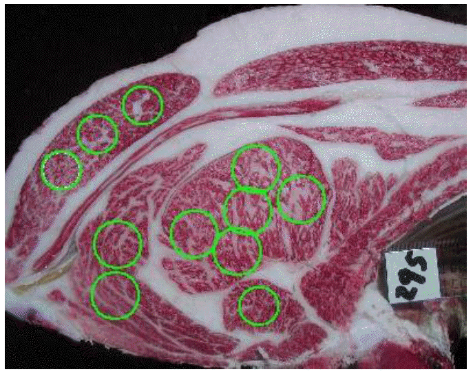
In the spot method, 3 to 5 locations (3.0 cm in diameter) for longissimus thoracis muscle, 3 locations 2.5 cm in diameter for trapezius muscle, 2 locations 3.0 cm in diameter for semispinalis dorsi muscle and 1 location 2.5 cm in diameter for semispinalis capitis muscle were selected rapidly on the monitor (Figure 2). The images were flattened, binarized and measured for ratio of fat area to muscle area by the software. In the spot method, there was no additional manual trace.
RESULTS AND DISCUSSION
Measurements by image analysis
Table 1 shows the average, standard deviation, maximum and minimum values by image analysis. The measurements at the same muscles were similar between the methods. The slight differences in values by methods at the same muscle may be caused by slight differences in automatic treatment of image analysis and/or in marbling distribution between the surrounding and the center parts in muscle. But, this will not actually become a problem as shown the following.
Correlation of the area and marbling between each muscle
Table 2 shows the correlation coefficients between muscle areas. All correlation coefficients between muscle areas were above 0.5. The areas of the longissimus thoracis muscle in particular were relatively strongly correlated with semispinalis dorsi muscle (r = 0.81), semispinalis capitis muscle (r = 0.72) and trapezius muscle (r = 0.63) (p>0.01).
These results indicate that the measurement of parameters of 1 muscle area, especially the longissimus thoracis muscle, can indicate the values for the other muscle areas. As the muscle area relates with the volume of meat, the formulas for determining beef yield grade in USA (AMSA) and Japan (JMGA) incorporate the area of the longissimus thoracis muscle.
Image analysis can measure longissimus muscle area and predict yield grade (Karnuah et al., 2001; Steiner et al., 2003). Determination of longissimus muscle area is fast and can be automate for lean beef (Steiner et al., 2003) but requires manual operation for highly marbled beef (Nade et al., 2004). Nade et al. (2004) reported that a new equation incorporating total muscle areas in the loin by image analysis is more suitable for Japanese beef yield grade than the present equation containing the longissimus muscle area only. A rough estimation of all muscle areas in the loin is quicker than an accurate measurement of each muscle area manually for yield grade of marbled beef.
Table 3 displays the correlation coefficients of marbling between muscles. In the overall trace method for marbling, the marbling in the longissimus thoracis muscle was relatively strongly correlated with semispinalis dorsi muscle (r = 0.73), semispinalis capitis muscle (r = 0.69) and trapezius muscle (r = 0.70) (p>0.01). Marbling in the loin was similar high correlation coefficient to muscle area. Although the marbling of 1 muscle was comparatively highly related with marbling of the other muscles in loin, the relationships may not always apply for other portions such as the hip (unpublished data). Meat packers in Japan gaze the marbling in other portions such as the hip, in addition to marbling in the loin.
It was reported that fat area determined by image analysis related with lipid content (r = 0.71, Yang et al., 2006; r = 0.98, Kuchida et al., 2000). Measurements of marbling by image analysis at meat markets can provide information on lipid contents and calories to the consumer.
Relationships between the simple spot and overall trace methods
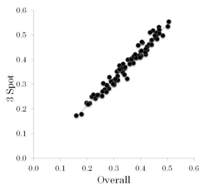
Table 2 also shows that the relationships between the overall trace and spot methods. The spot method for beef marbling related strongly with the overall trace method (from minimum value r = 0.89 to maximum value r = 0.97) in all muscles. These results showed that the spot method can substitute the overall trace method. As an example, the relationship between the measurements by overall trace and 3 spot methods for longissimus thoracis is shown in Figure 3. These data are to be comparatively fitted by a straight line. Although the spot method cannot determine muscle area, it has some merits. The spot method does not entail the time-consuming manual operation of the overall method. Even when the overall image is not obtained because of a narrow cut, the spot method can make use of even a partial image. Because the constant area is measured, the spot method is used to measure only the fat area, thus measuring of 2 or more spots do not take time. This enables the miniaturization of the photographic device with constant illumination by a spot light, such as fiber-optic devices (Irie and Swatland, 1993). In addition, this method can be applied to other rounded parts such as the hip. Hip is second important parts for marbling evaluation. Some beef carcasses with high marbling at loin have occasionally low marbling at hip. When this method is combined with the CT image (Holl├│ et al., 2007), the evaluation of the marbling on various tissues might speed up. Further study including new capture devices for spot method is needed for application to meat market.
 PDF Links
 PubReader
 ePub Link
 Full text via DOI
 Full text via PMC
 Download Citation
 Print
Print


