Effects of Chitosan on Body Weight Gain, Growth Hormone and Intestinal Morphology in Weaned Pigs
Article information
Abstract
The study was conducted to determine the effects of chitosan on the concentrations of GH and IGF-I in serum and small intestinal morphological structure of piglets, in order to evaluate the regulating action of chitosan on weaned pig growth through endocrine and intestinal morphological approaches. A total of 180 weaned pigs (35 d of age; 11.56±1.61 kg of body weight) were selected and assigned randomly to 5 dietary treatments, including 1 basal diet (control) and 4 diets with chitosan supplementation (100, 500, 1,000 and 2,000 mg/kg, respectively). Each treatment contained six replicate pens with six pigs per pen. The experiment lasted for 28 d. The results showed that the average body weight gain (BWG) of pigs was improved quadratically by dietary chitosan during the former 14 d and the later 14 d after weaned (p<0.05). Furthermore, dietary supplementation of chitosan tended to quadratically increase the concentration of serum GH on d 14 (p = 0.082) and 28 (p = 0.087). Diets supplemented with increasing levels of chitosan increased quadratically the villus height of jejunum and ileum on d 14 (p = 0.089, p<0.01) and 28 (p = 0.074, p<0.01), meanwhile, chitosan increased quadratically the ratio of villus height to crypt depth in duodenum, jejunum and ileum on d 14 (p<0.05, p = 0.055, p<0.01) and 28 (p<0.01, p<0.01, p<0.01), however, it decreased quadratically crypt depth in ileum on d 14 (p<0.05) and that in duodenum, jejunum and ileum on d 28 (p<0.01, p<0.05, p<0.05). In conclusion, these results indicated that chitosan could quadratically improve growth in weaned pigs, and the underlying mechanism may due to the increase of the serum GH concentration and improvement of the small intestines morphological structure.
INTRODUCTION
Chitosan, a deacetylated chitin, is widespread in nature. The exoskeletons of arthropods such as crabs, shrimps, insects, and other marine creatures in the crustacean family are good sources of chitosan (Crini, 2005; Huang et al., 2007; Li et al., 2009). Chitosan is a natural alkaline polysaccharide with positive charges, and also one of the most abundant natural polymers (Knaul et al., 1999; Wang and Zhang, 2004; Xu and Wang, 2005). As a nontoxic, biodegradable carbohydrate polymer (Goiri et al., 2010), it contains amino and hydroxyl groups per residue (Liao et al., 2007), which give chitosan many biological activities, such as haemostatic (Pusateri et al., 2006), anti-inflammatory (Dai et al., 2009), antitumor activity (Tsukada et al., 1990; Koide, 1998), antimicrobial activity (Limam et al., 2011; Benhabiles et al., 2012), hypoglycemic and hypocholesterolemic activity (Yao et al., 2006, 2008) and an immune-stimulatory effect (Moon et al., 2007; Yin et al., 2008).
Chitosan has become a new candidate as a growth-promoter for farm animals. Previous experiments involving piglets and broilers have demonstrated that chitosan improves growth performance of animals (Huang et al., 2005; Shi et al., 2005; Khambualai et al., 2009; Yuan and Chen, 2012). But the mechanism was not understood completely. Therefore, the present experiment was conducted to determine the effects of dietary chitosan supplementation on serum GH and IGF-1 and small intestinal morphological structure in weaned pigs.
MATERIALS AND METHODS
The protocol of the present experiment was approved by the Animal Care and Use Committee, Inner Mongolia Agricultural University, Huhhot, China.
Animals and experimental design
A total of 180 crossbred pigs (Duroc×Yorkshire ×Landrace; average body weight of 11.56±1.61 kg; 35 d of age) were used in this study. The pigs were weaned at 28 d of age. After weaning, the pigs were moved into the nursery building and given a 7-d adjustment period to adapt to the building and the dietary treatment. Pigs were randomly allotted to five treatments based on body weight and gender in a single factorial arrangement. Each treatment had 6 replicates (three replicate pens of males and three replicate pens of females) with 6 pigs per replicate. The five treatments were a basal diet (control group) and 4 diets with chitosan supplementation (100, 500, 1,000 and 2,000 mg/kg, respectively).
The basal diet met or exceeded nutrient requirements recommended by the NRC (1998) for swine and contained no chitosan (Table 1). The chitosan used in this experiment was provided by Jinan Haidebei Marine Bioengineering Co, Ltd. (Shandong Province, China) and its degree of deacetylation was determined to be 85.09%, and viscosity to be 45 cps.
The pigs were housed in a temperature controlled room and plastic floored pens of 4.00×4.20 m2 size with a self-feeder and nipple drinker to allow ad libitum access to feed and water. The environmental temperature was initially established at 28°C, which gradually decline to 20°C by the end of the experiment.
Sampling and measurements
Growth performance:
The pigs were individually weighed at the start and on d 14 and 28 of the trial, and the average body weight gain (BWG) was calculated.
Blood sample:
On d 14 and 28, peripheral blood was obtained through puncture of vena cava from one pig per pen and collected in a 5-mL evacuated tube (Becton Dickinson Vacutainer Systems, Franklin Lakes, NJ). Immediately after blood collection, the tubes were gently rocked several times and centrifuged (1,200×g for 15 min). After centrifugation, serum samples were labeled and stored at −80°C in cryogenic tubes until concentrations of GH and IGF-I were analyzed. The concentrations of GH and IGF-I were analyzed using commercially available RIA kits (Beijing Huaying Institute of Biological Technology, Beijing, China). All steps of the RIA for the serum GH and IGF-I were performed according to the kit protocol.
Small intestinal morphological structure:
On d 14 and 28 of the experimental period, one pig was randomly selected from each pen and killed by intravenous injection of sodium pentobarbital. The gastrointestinal tracts were quickly removed, and approximately 2 cm segments of the duodenum, jejunum and ileum were taken from the middle of each part, cut transversely, washed with physiological saline and covered with tissue-tek O.C.T (Sakura finetek Japan Co., Ltd., Tokyo, Japan). The segments were cut approximately 7 μm thick with a freezing microtome (Leica Microsystems Co., Ltd., Shanghai, China), and stained with haematoxylin and eosin (Nabuurs et al., 1993). A total of ten intact, well-oriented crypt-villus units were selected for each intestinal cross-section. The villus height and the crypt depth were measured using an image processing and analysis system (Version 1, Leica Imaging Systems Co., Ltd., Cambridge, UK), and then the ratio of villus height to crypt depth (VH/CD) was calculated. Villus height was measured from the tip of the villus to villus-crypt junction and crypt depth was defined as the depth of the invagination between adjacent villi (Hou et al., 2006).
Statistical analysis
All experimental data were analyzed in accordance with the GLM Procedure established by the SAS Institutes (2003). Regression analysis was conducted to evaluate linear and quadratic effects of chitosan levels on the various parameters measured in this experiment. Experimental unit was the replicate for all analysis. The BWG data were further analyzed by broken-line analysis to determine the optimal level of chitosan supplementation (Liu et al., 2008). The variability of the data was expressed as the standard error and a probability level of p<0.05 was considered to be statistically significant, whereas a p<0.10 was considered to constitute a tendency.
RESULTS
Growth performance
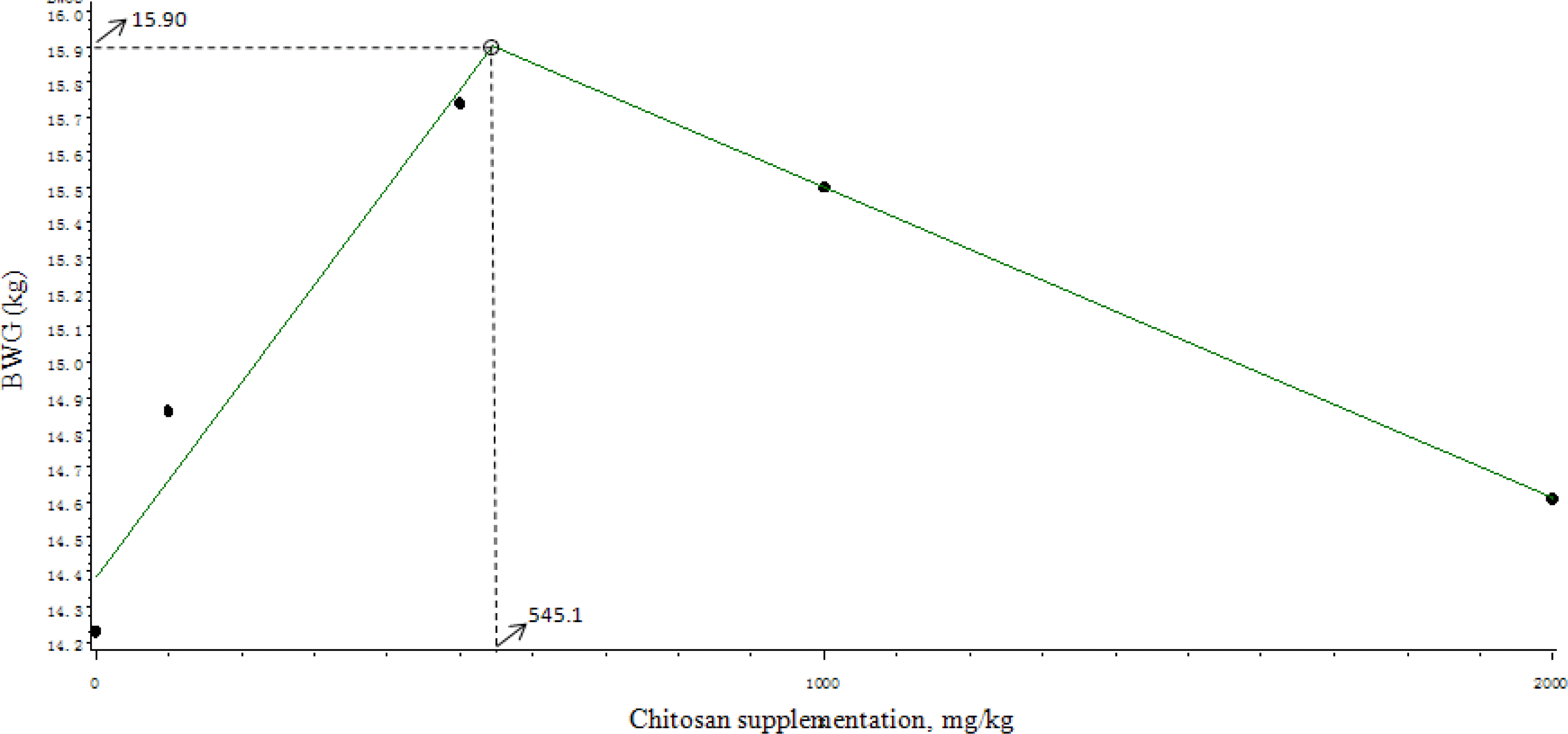
Effects of chitosan supplementation on growth performance of weaned pigs are shown in Table 2. Dietary supplementation of chitosan had a quadratic improving tendency (p = 0.064) on final body weight on d 14 and quadratic improving (p<0.05) on d 28. Furthermore, dietary chitosan quadratically improved average body weight gains in the first 14 d (p<0.05) and the second 14 d (p<0.05). The broken-line analysis on BWG of pigs during the entire study period of 28 d indicated that the optimal level of chitosan supplementation was 545.1 mg/kg (Figure 1).

Broken-line analysis of BWG of pigs given diets with different levels of chitosan supplementation during the entire study period of 28 d. The broken-line analysis indicated the breakpoint as 545.1 mg/kg, showing that the maximal BWG can be obtained by supplementation of 545.1 mg of chitosan/kg (p = 0.006, R2 = 0.377).
Serum GH and IGF-I
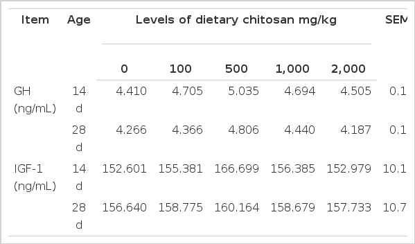
Effects of chitosan supplementation on serum GH and IGF-I of weaned pigs are shown in Table 3. Dietary supplementation of chitosan tended to quadratically increase the concentration of serum GH on d 14 (p = 0.082) and 28 (p = 0.087). However, there was no effect on serum IGF-1 with increasing chitosan supplementation.
Small intestinal morphological structure
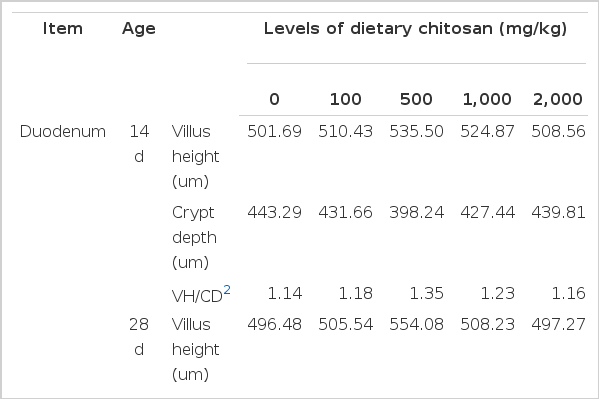
Effects of chitosan supplementation on small intestinal morphological structure of weaned pigs are shown in Table 4. Dietary chitosan quadratically increased the ratio of villus height to crypt depth in duodenum on d 14 (p<0.05) and 28 (p<0.01), meanwhile, quadratically decreased (p<0.01) crypt depth in duodenum on d 28. Diets supplemented with increasing levels of chitosan increased quadratically the villus height of jejunum on d 14 (p = 0.089) and 28 (p = 0.074), and the ratio of villus height to crypt depth on d 14 (p = 0.055) and 28 (p<0.01), however, decreased linearly (p<0.05) or quadratically (p<0.01) the crypt depth on d 28. In addition, dietary chitosan quadratically increased the villus height and the ratio of villus height to crypt depth in ileum on d 14 (p<0.01, p<0.01) and 28 (p<0.01, p<0.01), however, linearly (p< 0.05) and quadratically (p<0.05) decreased crypt depth on d 14 and quadratically (p<0.05) decreased crypt depth on d 28.
DISCUSSION
The weaning period is one of the most stressful phases after birth with weaning process inducing digestive disorder, intestinal barrier dysfunctions and impaired performance (Smith et al., 2010; Peace et al., 2011; Kim et al., 2012). An improvement in pig growth performance for economic purposes can be achieved by enhancing growth rate. One of the strategies to reduce weanling stress is to provide a specific feed additive, such as chitosan. In the current study, chitosan was effective in increasing weight gain of weaned pigs, which was in agreement with established literature (Huang et al., 2005; Shi et al., 2005; Khambualai et al., 2009; Yuan and Chen, 2012).
Growth in pigs is regulated in large part by the brain neuroendocrine GH-IGFs axis (Hall et al., 1986). GH can influence skeletal and muscle growth via direct and indirect effects on protein, lipid and carbohydrate metabolism (Pell and Bates, 1990) and fulfills its function of growth promotion through improving the production of IGF-I. IGF-I can stimulate amino acid uptake and protein synthesis in muscles and can greatly reduce the rate of protein breakdown within muscle fibers (Liu et al., 2008). Tang et al. (2005) reported that dietary supplementation of chitosan increased the serum GH and IGF-I levels. As shown in present study, the weaned piglets fed the diets supplemented with increasing levels of chitosan had a quadratic increase tendency in serum GH concentration compared with the control group, but there was no difference in the concentration of serum IGF-1 among treatments. The causes of this result are unclear and requires further experimentation to solve.
The small intestine is the main place for digestion and absorption of nutrients, and the intestinal mucosa plays an important role in these processes. Weanling stress can result in relatively quick changes in the intestinal mucosa morphological structure, which lead to a reduction in villus height and an increase in crypt depth (Hampson, 1986; Pluske et al., 1996a, b). Abnormal intestinal morphological structure is usually associated with retarded growth of weanling piglets. A shortening of the villus decreases the surface area for nutrient absorption, which lead to poor nutrient absorption and reduced performance (Xu et al., 2003). The crypt is the area where stem cells divide to permit the renewal of the villus, and a large crypt indicates fast tissue turnover and a high demand for new tissue (Hu et al., 2012). The ratio of villus height to crypt depth is a useful criterion for estimating the digestive capacity in the small intestine (Montagne et al., 2003). In agreement with the improved performance in weanling piglets, chitosan resulted in an improvement of intestinal morphological structure, as indicated by the increased small intestinal villus height and the ratio of villus height to crypt depth, and the decreased small intestinal crypt depth of weaned pigs in this study. Similar results were also reported by other investigators (Torzsas et al., 1996; Han et al., 2012). It is well known that pathogenic germs such as coliforms can destroy the normal morphology of small intestinal mucosa. Our previous study indicated that dietary chitosan could inhibit the proliferation of E. coli in the intestine, and improve gut microecology (Xu et al., 2012). Furthermore, chitosan provided a beneficial environment for the proliferation of enterocytes, preventing intestinal atrophy (Han et al., 2012). These studies showed that chitosan was an effective polysaccharide in ameliorating intestinal structure and function, which may be one of the reasons for the increased growth performance in weaned pigs treated with chitosan.
From the results of this study, it can be concluded that chitosan supplementation in diets of weaned pigs is helpful in improving growth rate, and the improvement mechanism may be partly attributed to increased GH concentration in serum and ameliorated morphological structure of small intestine. The optimal response occurred at 500 mg/ kg.
Acknowledgements
This research was supported by National Natural Science Foundation, China (Project No. 31060310).


