Effect of Feeding Bacillus subtilis natto on Hindgut Fermentation and Microbiota of Holstein Dairy Cows
Article information
Abstract
The effect of Bacillus subtilis natto on hindgut fermentation and microbiota of early lactation Holstein dairy cows was investigated in this study. Thirty-six Holstein dairy cows in early lactation were randomly allocated to three groups: no B. subtilis natto as the control group, B. subtilis natto with 0.5×10¹¹ cfu as DMF1 group and B. subtilis natto with 1.0×10¹¹ cfu as DMF2 group. After 14 days of adaptation period, the formal experiment was started and lasted for 63 days. Fecal samples were collected directly from the rectum of each animal on the morning at the end of eighth week and placed into sterile plastic bags. The pH, NH3-N and VFA concentration were determined and fecal bacteria DNA was extracted and analyzed by DGGE. The results showed that the addition of B. subtilus natto at either treatment level resulted in a decrease in fecal NH3-N concentration but had no effect on fecal pH and VFA. The DGGE profile revealed that B. subtilis natto affected the population of fecal bacteria. The diversity index of Shannon-Wiener in DFM1 decreased significantly compared to the control. Fecal Alistipes sp., Clostridium sp., Roseospira sp., beta proteobacterium were decreased and Bifidobacterium was increased after supplementing with B. subtilis natto. This study demonstrated that B. subtilis natto had a tendency to change fecal microbiota balance.
INTRODUCTION
Probiotics are living microbes which administered in adequate amounts confer a health benefit to the host (FAO/WHO, 2001). Some researches showed that probiotics can improve the performance of calves (Meyer et al., 2001; Timmerman et al., 2005), modify intestinal balance (Fuller, 1989) and mitigate calf scours (Wehnes et al., 2009). Probiotics can have several modes of action. These include - restricting colonization of pathogenic microbes on mucosal surfaces by a mechanism of competitive exclusion, stimulating an immune response, facilitating the proliferation of other commensal bacteria and producing antimicrobial substances (La Ragione et al., 2001; Hong et al., 2005; Leser et al., 2009).
The major probiotic strains are Lactobacillus, Saccharomyces, Bacillus, Streptococcus, and Aspergillus (Tannock, 2001), and so on. Most of the previous research on probiotics were focused on the application of various strains of lactic acid bacteria (Miettinen et al., 1996; Holzapfel et al., 2001). Several Bacillus sp. bacteria, such as Bacillus licheniformis and Bacillus subtilis have gained more attention for their role in controlling infectious diseases, thereby improving productive performance in animals (Holzapfel et al., 2001). Unlike the lactic acid bacteria, Bacillus sp. is not normally found in the gastrointestinal tract. Bacillus sp. exists in an endosymbiotic relationship with their host, being temporarily able to survive and proliferate within gastrointestinal tract (GIT) and finally excreted in the faeces (Hong et al., 2005). Bacillus sp. also provides beneficial effects via one or a number of the modes of action described above (Wehnes et al., 2009). Bacillus sp. have demonstrated effectiveness as probiotics because they have been shown to be capable of inhibiting pathogens, such as Clostridium sp. (Guo et al., 2006), Campylobacter sp. (Fritts et al., 2000), Streptococcus sp. (Teo and Tan, 2005), Escherichia coli (Teo and Tan, 2006), Salmonella typhimurium, and Staphylococcus aureus (Sumi et al., 1997). B. subtilis natto isolated from the Japanese fermented soybean staple known as ‘natto’ is a gram-positive spore-forming bacterium and is a subspecies of B. subtilis according to the 16S rRNA sequence analysis (Qin et al., 2005). Our prior research suggested that the viable probiotic characteristics of B. subtilis natto was beneficial to the immune system of the calf (Sun et al., 2010).
The gastrointestinal tract of healthy animals is colonized by a complex microbiota formed by many different species of microorganism (Frizzo et al., 2008). The microbial balance in the microbiota of the digestive tract is important to promote efficient digestion and maximum nutrient absorption. It can increase the host capacity for excluding pathogen microorganisms and thus, to prevent some diseases at the same time (Walter et al., 2003). The microbial shedding and fermentation parameters of the feces can particularly reflect the true condition of the hindgut (Fox et al., 2007). However, the effects of dietary supplementation with B. subtilus natto on hindgut microbiology and microbial population have been rarely investigated. Consequently, this study was designed to assess the effect of B. subtilis natto on hindgut microbiota and fermentation of Holstein dairy cows.
MATERIALS AND METHODS
Cows and diets
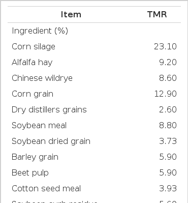
All animals used in this experiment were maintained according to the principles of the Chinese Academy of Agricultural Sciences Animal Care and Use Committee. The study was conducted at the Beijing Dairy Cattle center in Yanqing Conty, Beijing. Thirty-six early lactation Holstein dairy cows were used in this experiment, and the experiment dates were from March 9th to May 3rd, 2010. Cows were fed a typical total mixed ration (TMR) based on China Standard NY/t 34 (2004) and NRC (2001) (Table 1) and pre-weighted rations (110%) of TMR was supplied to the cows every meal.
Experimental design
After two weeks of adaptation feeding, cows were divided into three groups: i) no B. subtilis natto as the control group (CON); ii) cows were fed the TMR diet supplemented with 6 g/d B. subtilis natto fermentation product (0.5×10¹¹ cfu of B. subtilis natto/d) (DFM1); and iii) cows were fed the TMR diet supplemented with 12 g/d B. subtilis natto fermentation product (1.0×10¹¹ cfu of B. subtilis natto/d) (DFM2). The TMR was given three times a day at 0700, 1400 and 2100 with the B. subtilis natto being added daily in the morning feeding. The solid state fermentation products were produced by Langfang Dongxin Biological Technology Co., LTD (China) using the prior identified B. subtilis natto from our laboratory. The number of viable B. subtilis natto spores in the product was determined by plate count on LB medium (5% agar, 0.5% NaCl, 1% soya peptone, 0.3% beef extract, pH = 7.0) after heat treatment (80°C for 10 min), and approximately 8.3×10⁹ spores per gram of solid-state fermentation product were obtained. Animals had free access to fresh water and a plain mineral block during the period of the experiment.
Sample collection and analysis
Fecal samples were collected directly from the rectum of each animal on the morning at the end of eighth week and placed into sterile plastic bags. These bags were kept on ice until the sampling of all animals was completed. The entire process was limited to one hour’s duration
Fecal pH
The pH of fresh feces was determined immediately after defecation by thoroughly mixing 10 g fresh feces with 20 mL double-deionized water in 50 mL tubes and submerging the pH probe (370 model pH meter, Jenway, UK) in the mixture (Breg et al., 2005). Then 4ml of the feces suspension was placed into a 10 mL sterile test tube. 1 mL 25% meta-phosphoric acid was added into 4 mL of the feces suspension, then mixed well and stored at −20°C for further NH3-N and VFA analysis.
Fecal NH3-N and VFA
Fecal NH3-N was measured according to Bromner and Keeney (1965). Fecal VFA concentrations were determined using gas chromatography (model 6890, Series II; Hewlett Packard Co., Avondale, PA) using a DB-FFAP (15 m×0.32 mm×0.25 μm) and FID. The samples were run at a split ratio of 50:1 with a programmed temperature gradient (100°C initial temperature for 1 min, with a 2°C rise per min to 120°C and a 10 min final temperature). The temperature of the injector and detector was 250°C and 280°C respectively. The carrier gas was N2, and column flow rate was 1 mL/min (Mohammed et al., 2004).
PCR-DGGE
The fecal DNA of the eight week was extracted by the method of RBB+C (Yu et al., 2004). For DGGE analysis, approximately 200 bp of the fecal total 16S rDNA gene was amplified using primers: 338f (5′-ACTCCTACGGGAGGCAGC AG-3′) and 533r (5′-TTACCGCGGCTGCTGGCAC-3′) and with a 40-base GC-clamp (CGCCCGCCGCGCGCGGCGGGCGGGGCGGGGGCACGGGGGG) at the 5′ terminus of 338f primer. All PCR amplifications were performed in 50 μL volumes containing 25 μL 2×HiFiTaq StarMix (GenStar, USA), 1 μL 500 nM each primer, 100 ng DNA template. After prior denaturation at 94°C for 4 min, 10 cycles of touchdown PCR were performed (94°C for 30 s, 61°C for 30 s, with a 0.5°C per cycle decrement, and 72°C for 1 min), followed by 25 cycles of PCR (94°C for 30 s, 56°C for 30 s, and 72°C for 1 min), and a final extension step at 72°C for 7 min.
The PCR-DGGE was performed using a DCode Universal Mutation Detection System (Bio-Rad Laboratories, Hercules, CA, USA). Fifteen μL of PCR products were loaded in a 7.5% polyacrylamide/gel (37.5:1) in 0.5×TAE (20 mmol/L Tris-HCl, 10 mmol/L acetic acid, 0.5 mmol/L EDTA adjusted to pH 8.3) buffer. The polyacrylamide gels contained a 40% of denaturant at the top of the gel grading to a 60% denaturant at the bottom (100% denaturants consisting of 40% [v/v] formamide and 7 M urea). The DGGE gel was run for 16 h at 60°C and 85 V. After electrophoresis, the gels were stained with silver nitrate (Yu et al., 2004).
Sequencing
Putative indicator bands were excised from the gels and DNAs were recovered according to boiling methods (Wang et al., 2006). The 16S rDNA from each band was enriched by PCR using the same primer 338f and 533r without a GC-clamp under the prior PCR process. Purified PCR products were cloned into pMD18-T vector, and transformed into Escherichia coli JM109 competent cells following the manufacturer’s instructions (TaKaRa, Japan). Screening the positive clones in the LB solid medium containing X-Gal and ampicillin (Amp), and the sequencing was completed by the BGI Company, Beijing center (Beijing, China). After aligning with sequences from GenBank and RDP, 16S rDNA sequences were blasted individually with the highest similar sequence.
Statistical analysis
All data were analyzed using the Proc Mixed procedure of the SAS system (version 8.2, SAS Institute Inc., Cary, NC). p-Values <0.05 were considered statistically significant, and trends were discussed at p<0.1.
For microbial diversity analysis, after staining, DGGE gels were scanned and analyzed using Quantity-one software (Bio-Rad) and the peak density was calculated. The unweighted pair group method using arithmetic averages (UPGMA) algorithm was used as implemented in the analysis software for the construction of dendrograms. The microbial diversity was analyzed according to Shannon-Wiener index (H′) (Spellerberg and Fedor, 2003):
H′ = −∑Pi logPi
Pi = Ni/N, relative intensity in the profile
Ni = surface of the peak i
N = sum of the surfaces for all the peak within the profile
RESULTS
Fecal pH, NH3-N and VFA
All results are listed in Table 2. There were no treatment effects on fecal pH during this study. Compared to the Control group, supplementation of B. subtilis natto in the diets led to a significant decrease in total fecal NH3-N in DFM1 and DFM2 (p<0.10). No differences were observed in total fecal VFAs or the proportion of individual VFAs such as: acetate, propionate, butyric, isobutyric, valeric or isovaleric when cows were supplemented with either 0.5×10¹¹ cfu of B. subtilis natto/d or 1.0×10¹¹ cfu of B. subtilis natto/d.
Fecal microbial analysis
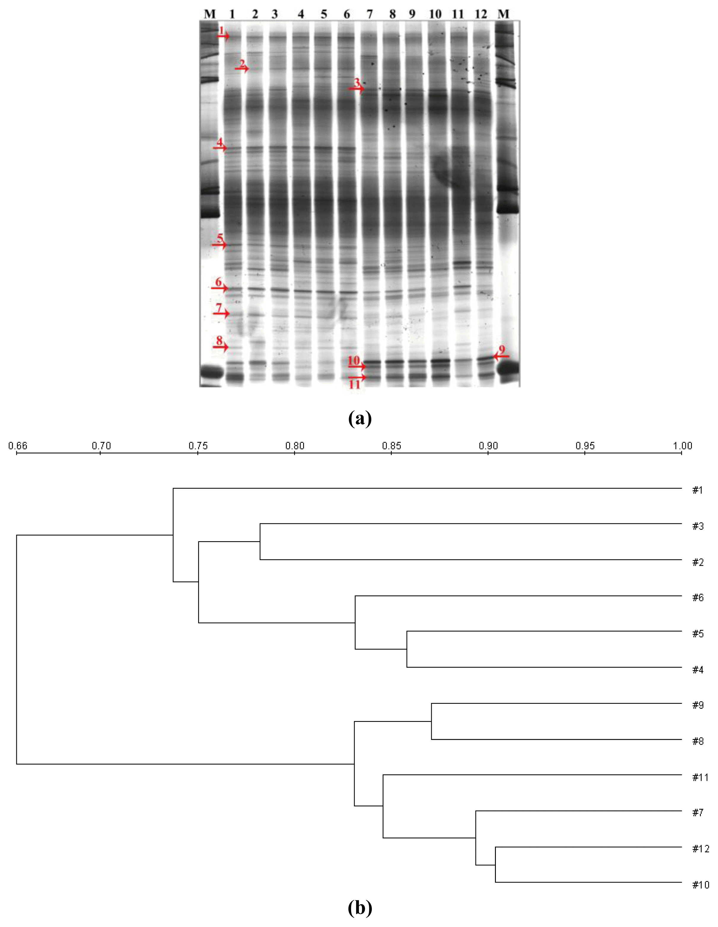
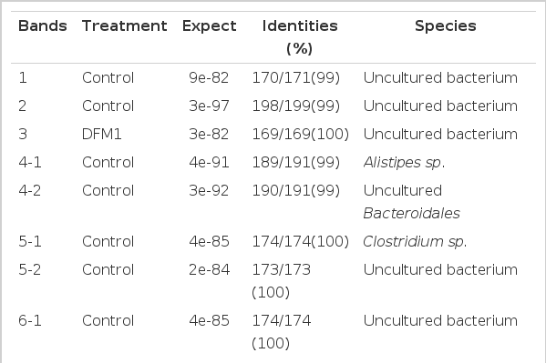
The total fecal DNA, DGGE fingerprint for the eighth week for both CON and DFM1 is shown in Figure 1(a). Examination of the gel indicated that the position and number of bands were similar in each group. However Bands 1, 2, 4, 5, 6, 7, and 8 were decreased in intensity or disappeared in the same fingerprint of the DFM1 group, while bands 3, 9, and 11 increased in intensity compared with the CON. In addition the band group labeled ‘10’ was a new band group which was not present in the CON fingerprint. It was also observed that there were numerous color differences in this gel that were implied the difference in density. Dendrograms shown that lanes belonged to the same group: lanes 1, 2, 3, 4, 5, 6, and lanes 7, 8, 9, 10, 11, 12 clustered respectively and the similarity of the two groups was about 66% (Figure 1(b)). Labeled bands were excised and sequenced, the results are shown in Table 3, fecal Alistipes sp., Clostridium sp., Roseospira sp., beta proteobacterium were decreased and Bifidobacterum was increased after supplementing B. subtilis natto. However, some bands were changed such as bands group 1, 2, 3, 7, and 11, that were uncultured bacteria sp.. Even if they were excised from the band groups, the sequence was inconsistent. For example − bands 4, 5, 6, 9, and 10 generated two different results. The Shannon-Wiener index of CON and DFM1 was 3.3476±0.07 and 3.0725±0.08 respectively, and a significant difference was obtained in our study when supplementing B. subtilis natto in the diets at eight week (p<0.10).

(a) DGGE profiles of 16S rDNA sequences amplified from DNA extracted from the eight week of the trial. Lane 1 to 6: CON; lane 7 to 12: DFM1. (b) UPGMA clustering of Figure 1(a). No. 1, 2, 3, 4, 5, 6 represent CON; No. 7, 8, 9, 10, 11, 12 represent DFM1.
DISCUSSION
This study investigated the effects of supplementing B. subtilis natto on hindgut fermentation and microbiota through fecal. No obvious changes in fecal pH were observed, however, a small decline in fecal pH was previously associated with altered fermentation patterns and microbial ecology in the hindgut (Medina et al., 2002). Similar results were also observed by Qiao et al. (2010). The decrease of fecal pH in this study may have contributed to the increase of Bi dobacteria in the hindgut of the cows fed B. subtilis natto. It is also possible that the decline of fecal pH may connect with the decrease of fecal NH3-N concentration. In this study — when the B. subtilis natto product was added to the diets — the concentration of total dietary N did not change. However, the resultant concentration of fecal NH3-N was decreased (p<0.01). Fecal N contains mainly undigested feed protein and metabolic fecal N (de Boer et al., 2002). Therefore, these results can be explained by assuming that the supplementation of B. subtilis natto product enhanced the diet utilization efficiency and reduced the excretion of protein in the feces (Spiehs et al., 2009). Previous research suggested that feeding fermented products of B. subtilis to layer hens (Santoso et al., 1999) and broilers (Santoso et al., 1995) reduced NH3-N release from excreta significantly, so we would surmise that there is a microbiological component which can reduce urease activity within the fecal microbial population. No difference in fecal VFAs or the concentration of individual VFAs was observed in the treatment groups that were supplemented with B. subtilis natto. Similar results were reported in meat goats, but no consistent benefit was noted from supplementing healthy, growing meat goats with probiotic products (Whieley et al., 2009).
The microbial population of the intestine plays an important role in overall animal health, productivity and well-being. Therefore, assessing the diversity of microbial communities (in terms of richness and structure) is a useful indicator as to how animals evolve within their environment (Fromin et al., 2002). In this study we reported the changes in hindgut microflora as measured in the faeces after supplementing B. subtilis natto using DGGE methods. There was a significant difference for the Shannon-Wiener index (H′) between the CON group and the DFM1 group at the eighth week, suggesting that the microbial population in the rumen of the cows in this study was affected by supplementing B. subtilis natto. Studies have demonstrated that probiotics can provide good production responses in various fields of animal production (Fuller, 1989; Hosoi et al., 2000; Hong et al., 2005; Frizzo et al., 2008). Probiotics can competitively exclude pathogenic bacteria or be antagonistic towards their growth, thus helping to maintain the health of the intestinal tract (Jin et al., 1997; Hong et al., 2005). In addition, probiotics affects the bacterial population and mix as measured in the faeces (Whitley et al., 2009). An increase in the level of Bifidobacterium sp. and a decrease in Alistipes sp., Clostridium sp., Roseospira sp. was found in this study. This is consistent with changes in Bifidobacterium and Clostridium sp. reported by Guo et al. (2006), E. coli 0157:H7 reported in lamb faeces by Lema et al. (2001) and a reduction in Salmonella shedding in beef cattle reported by Stephens et al. (2007). However inconsistent results were reported by other researchers where the abundance and population density of dairy cow were improved by the addition of B. subtilus Pab02 (Pan et al., 2010) but no effect was found in fecal microflora after supplementing the diets of meat goats with a probiotic (Whitley et al., 2009).
CONCLUSIONS
The addition of B. subtilus natto to the diets of dairy cows in early lactation was shown to significantly reduce NH3-N in the faeces indicating a possible improvement in nitrogen utilization. In addition B. subtilus natto may change the microfloral mix within the faeces. More research is required to understand the mode of action, and the effects of these changes on animal production.
ACKNOWLEDGMENTS
The investigation was financially supported by National Basic Research Program (973) of China (2011CB100805) and grants (No.2012BAD12B02-5 and No. 2004DA125184G1103) from Ministry of Science and Technology.


