The relationship between the variants in the 5′-untranslated regions of equine chorionic gonadotropin genes and serum equine chorionic gonadotropin levels
Article information
Abstract
Objective
An experiment was conducted to study the association between the single nucleotide polymorphisms (SNPs) in 5′-untranslated regions (5′-UTR) of equine chorionic gonadotropin (eCG) genes and the serum eCG levels.
Methods
SNPs in 5′-UTR of eCG genes were screened across 10 horse breeds, including 7 Chinese indigenous breeds and 3 imported breeds using iPLEX chemistry, and the association between the serum eCG levels of 174 pregnant Da’an mares and their serum eCG levels (determined with ELISA) was analyzed.
Results
Four SNPs were identified in the 5′-UTR of the eCGα gene, and one of them was unique in the indigenous breeds. There were 2 SNPs detected at the 5′ end of the eCGβ subunit gene, and one of them was only found in the Chinese breeds. The SNP g.39948246T>C at the 5′-UTR of eCGα was associated significantly with eCG levels of 75-day pregnant mare serum (p<0.05) in Da’an mares. Prediction analysis on binding sites of transcription factors showed that the g.39948246T>C mutation causes appearance of the specific binding site of hepatocyte nuclear factor 3 forkhead homolog 2 (HFH-2), which is a transcriptional repressor belonging to the forkhead protein family of transcription factors.
Conclusion
The SNP g.39948246T>C at the 5′-UTR of eCGα is associated with eCG levels of 75-day pregnant mare serum (p<0.05).
INTRODUCTION
Equine chorionic gonadotropin (eCG) is a gonadotropic hormone produced in the chorion of pregnant mares, previously known as pregnant mare’s serum gonadotropin, which was found in 40 to 130 days pregnant mare serum, first reported by Cole et al [1]. eCG is composed of two dissimilar subunits, alpha and beta subunit consisting of 96 and 149 amino acids, respectively. The alpha subunit (eCGα) is the same as its counterparts of all other glycoprotein hormones (equine luteinizing hormone, equine follicle-stimulating hormone, equine thyroid stimulating hormone). The beta subunit (eCGβ) is specific for eCG and is responsible for receptor binding specificity. The eCG activity and specificity is mainly determined by the beta subunit, and the alpha subunit also has a significant effect on biological activity of the hormone [2]. The use of eCG in follicular stimulation and superovulation has substantial effects on follicular development and corpus luteum function [3,4]. It stimulates the growth of the dominant follicle [5,6] and promotes the increase of circulating progesterone concentrations in the subsequently estrus cycle [6,7].
Breeds, maternal and paternal effects, and fetal genders are the main genetic factors which influence the eCG secretion. Previous studies showed that the breed factor has a great influence on the secretion level of eCG. It was reported that the serum eCG concentration of ponies and Mongolia horses was significantly higher than draft horses and Thoroughbreds [8–10]. The mutations of human and cattle CG were identified, which are associated with male infertility, recurrent miscarriage and superovulation traits [11–13]. But the mutations of eCG genes and their biofunctions have not been fully studied, and only a single nucleotide polymorphism (SNP) in the 5′-untranslated regions (5′-UTR) of eCGβ was identified in previous study [10]. The objective of the present study was to investigate SNPs in eCG genes and to evaluate the association between the polymorphisms and serum eCG levels.
MATERIALS AND METHODS
Sample collection and DNA extraction
A total of 417 blood samples were collected, which included 7 Chinese indigenous breeds and 3 imported breeds (Table 1). Da’an (DA), Inner Mongolia (MG), Elunchun (ELC), and Ningqiang (NQ) were distributed in the north of China, and Jinjiang (JJ), Changdu (CD), and Baise (BS) were located in southern regions of China. Generally, the northern horses were higher and stronger than the southern horses. MG and ELC have a closer genetic relationship than others. Genomic DNA was isolated from samples using phenol-chloroform method and preserved at −20°C [14]. The amount of DNA from each sample was determined by measuring A260/A280 value using NanoDrop 2000 (Thermo Scientific, Waltham, MA, USA) and further evaluated by agarose gel electrophoresis. All samples were obtained following the principles approved by the Animal Care and Use Committee of China Agricultural University.
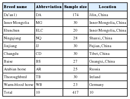
The location and breed information of the 417 samples
Single nucleotide polymorphism selection and genotyping
To detect SNPs, primers were designed using primer 5.0 software based on the sequences of the 5′UTRs of eCGα and eCGβ gene (Gene ID: 100034174 and 100054774). The primer pairs (eCGαF: 5′-CCCTTTCAATTTGTATGCC-3′, eCGαR: 5′-TATG CAGA AACACGGACAA-3′ and eCGβF: 5′-CAATGACTCGCTGAC CTCCTG-3′, eCGβR: 5′-GTCTCCATCCTCGGTGCCTC-3′) were designed to amplify the 1,818-bp and 1,266-bp fragments of 5′UTRs of eCGα and eCGβ gene, respectively. Polymerase chain reaction (PCR) amplifications were performed in a 20 μL reaction mixture consisting of the 25 ng DNA, 0.5 μM each primer, 2×Taq PCR MasterMix 10 μL (Tiange, Beijing, China) and ddH2O 8 μL. All amplifications included an initial denaturing step of 10 min at 95°C, followed by 35 cycles of 30 s at 95°C, 30 s at an optimized annealing temperature (eCGα, 54°C and eCGβ, 62°C) and 90 s at 72°C, with a final extension of 10 min at 72°C. The PCR products were sent to the Beijing Genomics Institute (Beijing, China) for sequencing.
The samples were genotyped by two steps. i) The polymorphisms in the 5′UTRs were screened by sequencing five individuals for each breed. ii) Based on the polymorphic information from the sequencing, all other samples were genotyped using iPLEX chemistry on a matrix-assisted laser desorption/ionization time-of-flight mass spectrometer (Applied Biosystems, Waltham, MA, USA).
Detection of eCG serum levels
Serum samples of 174 Da’an mare horses with ages between 4 to 11 years were collected three times on day 55, 65, and 75 of pregnancy, respectively. The eCG levels in serum were determined by enzyme-linked immunosorbent assay (ELISA) with commercially available kits (DRG, Marburg, Germany), according to the manufacturer’s instructions. The optical density (OD) at 450 nm was measured using an ELISA microplate reader (infiniteM200 NanoQuant)(TECAN, Hombrechtikon, Switzerland).
Statistical analysis
Genotypic and allelic frequencies for each SNP, Hardy–Weinberg equilibrium (HWE) x2 test for genotype distributions were analyzed using Microsoft Excel (Microsoft Corporation, Redmond, WA, USA). The genotype sample size and eCG serum levels of Da’an horses were analyzed by SPSS-Statistics V17.0 (Chicago, IL, USA). Association analysis between the SNPs and serum eCG levels of Da’an horses was analyzed following the Student-Newman-Keuls (SNK), yij = μ+ai+eij, where yij indicates observed value; μ, overall mean; ai, fixed effect of genotype; eij, random error. The binding sites of transcription factors were predicted using online software (http://www.phylofoot.org/) (Center for Genomics and Bioinformatics, Karolinska Institutet, Stockholm, Sweden).
RESULTS
SNPs in 5′ UTR of eCG genes
After sequencing and alignment, four SNPs in 5′UTR of eCGα gene (g.39947046C>G, g.39947259C>T, g.39947735C>G, g.399 48246T>C) were identified while two SNPs were found in the 5′UTR region of eCGβ gene (g.18962466G>T, g.18962516G>A).
The genotypes and allele frequencies of the SNPs in the 5′UTR of eCG genes are shown in Table 2, 3, 4. Results of x2 test indicated that the SNP g.39947046C>G and g.39947259C>T met the HWE (p>0.05) in all breeds, and g.39947735C>G and g.3994 8246T>C were in HWE only in the indigenous breeds.

Polymorphisms in the 5′ UTR of eCG genes
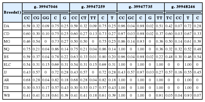
The allele and genotype frequencies for the polymorphisms of eCGα gene in the studied breeds
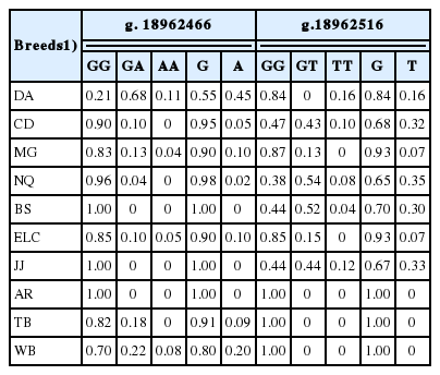
The allele and genotype frequencies for the polymorphisms of eCGβ gene in the studied breeds
Relationships between the SNPs of eCG genes and the serum eCG levels
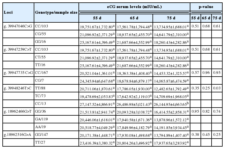
Effects of the 6 polymorphic loci of the eCG genes on the serum eCG levels on day 55, 65, and 75 of pregnancy of Da’an horses are shown in Table 5. Statistical analysis revealed that the eCG levels successively decreased with the increase of gestational time. SNP g.39948246T>C was associated with 75-day pregnant serum eCG levels (p<0.05), and there was more than twice as much average eCG concentration of individuals with CC genotype as that of horses harboring TT. Based on the transcription factors prediction, the mutation g.39948246T>C leads to the appearance of binding site of an important transcription factor, HFH-2.
DISCUSSION
The eCG genes are located on chromosome 10 in horse [15]. Reproductive physiology of eCGα and eCGβ subunits was subjected to intensive studies [2,16,17], while mutations in eCG genes have not been fully studied yet. Mutations of eCG gene may affect serum eCG levels. It was reported that a low level of human chorionic gonadotropin (hCG) in maternal serum during the first trimester of the pregnancy is a clinically accepted risk factor for miscarriage, and variation in hCGβ gene contributes to the susceptibility of recurrent miscarriage [12].
In this study, we detected 6 novel SNPs in eCGα and eCGβ genes by screening 10 horse breeds. Our results showed that the mutation g.39948246T>C in eCGα gene was significantly associated with the serum eCG levels, but its biofunction needs to be further studied. In Table 3, the indigenous and imported horse populations showed significant difference on allele frequencies at the mutant sites. And the imported horse breeds had the similar frequencies with each other, so did the indigenous breeds except ELC horses which experienced bottleneck of genetic diversity and presently has a small population. mRNA expression of eCG receptor indicated that eCG had high expression levels during 55 to 70 days of gestation [18]. As the results show in Table 5, g.39948246T>C was associated significantly with the serum eCG levels on day 75 of pregnancy (p<0.05). Due to effect of a gene-substitution, the horses harboring CC have higher eCG secretion levels. Therefore, the SNP g.39948246T>C may have the potential to be used as a genetic maker for the selection of high level of serum eCG.
Transcription factors play important role on controlling the flow of genetic information from DNA to mRNA [19]. According to the prediction results, at the mutant site of g.39948246T>C of eCGα, the substitution of T for C leads to the appearance of the binding site of the transcription factor HFH-2. Previous studies showed that HFH-2 (alias Foxd3) decreased interleukin-10 expression in B cells, and FOXD3/miR-214/MED19 axis suppresses tumors growth and metastasis in human colorectal cancer [20,21]. Individuals carrying TT genotype had lower eCG levels than those of GG genotype which may be attributed to the suppression caused by inhibitory effect of HFH-2.
CONCLUSION
In conclusion, we detected 6 SNPs in 5′ UTRs of eCGα and eCGβ genes. The SNP g.39948246T>C was significantly associated with eCG secretion levels, and individuals harboring genotype CC had the highest serum eCG level in all of the three genotypes. Thus, this SNP has the potential to be used as a molecular marker on the selection for high serum eCG levels. So the present study contributes new information on understanding functions of the eCG genes.
ACKNOWLEDGMENTS
The present study was financially supported by the Public Science and Technology Research Funds Projects of Agriculture (Grant No. 201003075), the Program for Changjiang Scholars and Innovative Research Team in University (Grant No. IRT_15R62), and the fund from Beijing Key Laboratory for Genetic Improvement of Livestock and Poultry.
Notes
CONFLICT OF INTEREST
We certify that there is no conflict of interest with any financial organization regarding the material discussed in the manuscript.