Effect of Monochromic Light-emitting Diode Light with Different Color on the Growth and Reproductive Performances of Breeder Geese
Article information
Abstract
The purpose of this study was to investigate the effect of monochromic light-emitting diode (LED) light with different color on the growth and reproductive performances of white Roman breeder geese. A randomized complete batch design was utilized for the trial, and the replicate was regarded as one batch. Twenty ganders and fifty-five dames were used in batch 1 (started on 2011/6/17 and ended on 2012/1/31), thirty ganders and eighty-four dames were used in batch 2 (started on 2012/3/23 and ended on 2012/10/26), and thirty ganders and seventy-two dames were used in batch 3 (started on 2013/3/12 and ended on 2013/12/20). Two hundred and ninety-one geese were randomly assigned to 6 rooms in an environmentally controlled house. They were randomly allotted into one of three monochromatic light treatments: Blue, red, or white. The results showed that there was no significant difference in body weight among the three lighting groups at any point throughout the experimental period. However, compared to the blue light group, significantly more eggs were produced by the red and white light groups (p<0.05). Furthermore, the laying period of the red light group was significantly longer than that of other two groups (p<0.05). In conclusion, our results suggested that red LED-light has the best effect on reproductive performance (i.e. longer laying period and higher total eggs number) at 30 lux light intensity, and is therefore a better choice for the management of breeding geese than blue or white LED-light.
INTRODUCTION
Under natural light conditions, domestic geese show an obvious seasonal pattern of reproductive activity (Zeman et al., 1990). The photoperiod not only plays an important role in initiating egg production, but is also beneficial in sustaining persistent egg production (Gongruttananun and Guntapa, 2012).
Light plays a pivotal role in vision, the release of various hormones; it also increases the efficacy of important factors related to the behavior and reproduction of birds (Biyatmoko, 2014). Moreover, it also activates the development of reactive substances, such as enzymes and vitamins in embryos (Er et al., 2007). Evidently, the source, spectrum, intensity and regimen of artificial light supplementation are critical factors for the modern management of the poultry industry (Borille et al., 2013; Morrill et al., 2014; Lee et al., 2015; Wang et al., 2015).
Like the human eye, the chicken eye is capable of viewing a narrow part of the light spectrum, 380 to 760 nm (Rozenboim et al., 2012). In addition to the eyes, the extra-retinal photoreceptors (in the hypothalamus or other sites of the brain) are sensitive to different wavelengths and are involved in photostimulation (Lewis and Morris, 2000; Kram et al., 2010). Chickens are typical of most diurnal birds in possessing seven photoreceptor cell types: One rod and six cones (Kram et al., 2010). Tetrachromatic color vision is mediated by four types of single cones that are maximally responsive to violet, red, blue and green light, respectively (Bowmaker and Knowles, 1977). Nuboer et al. (1992) showed that a better way to present the relative luminance of a lamp for domestic fowl (‘galluminance’) is by multiplying the spectral power distribution of lamps. Prescott and Wathes (1999) reported that broilers are able to ‘see’ into the UVA and differ from that of humans, showing higher sensitivities between 380<Lambda<507 nm. Moreover, Prescott et al. (2003) proposed that a minimum dark period of 6 h duration should be used in cases of pecking damage and cannibalism impacting bird welfare.
Hsu et al. (1990) reported that light intensity of 0 to 40 lux showed no significantly effect on egg production in geese. Pyrzak et al. (1984) indicated that the geese under light intensity of 20 lux had a higher egg production than that of 50 lux. The effects of monochromatic light on the productivity and egg quality in hens have been reported in a previous study (Borille et al., 2013). They concluded the commercial layers (Institut de Sélection Animale [ISA] Brown) had the best egg production when exposed to red and white light-emitting diode (LED) and incandescent lamps. However, the feed intake, egg weight and egg quality were not influenced by the light sources. Conversely, Morrill et al. (2014) pointed out that under blue and green monochromatic illumination, chicken broilers exhibited up to 6.0% and 8.9% increases in final body weight, respectively, compared to chickens kept under red or white light. Mobarkey et al. (2013) showed that selective photostimulation under green light caused a significant delay in the onset of egg production of chickens compared to the use of red light. However, little is known about the effect of monochromic light of each different color on the growth and reproductive performances of breeder geese. The objective of this study, therefore, was to find the most suitable single monochromatic light, among the colors chosen in this study, during the laying period of white Roman geese.
MATERIALS AND METHODS
Animals and experiment design
A total of 80 ganders and 211 dames in their first laying season, with an average age of 10 months, were used as the objects of this study. Once the geese were moved into an environmentally controlled house, a restricted amount of resting rations (13.0% crude protein [CP] and 2,350 kcal/kg metabolizable energy [ME]) was provided according to the experimental protocol, and the photoperiod was adjusted to 7 L:17 D for the first 6 weeks. Thereafter, the photoperiod was adjusted to 9 L:15 D and the laying geese were fed ad libitum. The laying diet contained 18.0% CP and 2,700 kcal/kg ME. The care and use of all geese followed the guidelines set by the Regulations of Laboratory Animals, Changhua Animal Propagation Station, Livestock Research Institute, Council of Agriculture, Taiwan. Drinking water was provided ad libitum. A randomized complete batch design was utilized for testing, and the replicate was regarded as one batch. Twenty ganders and fifty-five dames were used in batch 1 (started on 2011/6/17 and ended on 2012/1/31), thirty ganders and eighty-four dames were used in batch 2 (started on 2012/3/23 and ended on 2012/10/26), and thirty ganders and seventy-two dames were used in batch 3 (started on 2013/3/12 and ended on 2013/12/20). In total, each light spectrum involved 6 repeats/pens (included 3 batches). Each pen was 2.4 m×6.5 m (15.6 m2), signifying 0.77 bird per m2 in batch 1, 1.22 bird per m2 in batch 2 and 1.09 bird per m2 in batch 3. The randomized complete design included three monochromatic lights (LED lights), i.e. blue, red and white light, for a total of 3 groups at the Changhua Animal Propagation Station (23°51′N, 120°33′E), Taiwan (Figure 1). For focusing on the effect of light color, the light intensity was fixed at 30 lux for all the 3 light colors at the height of geese eyes when they were standing.
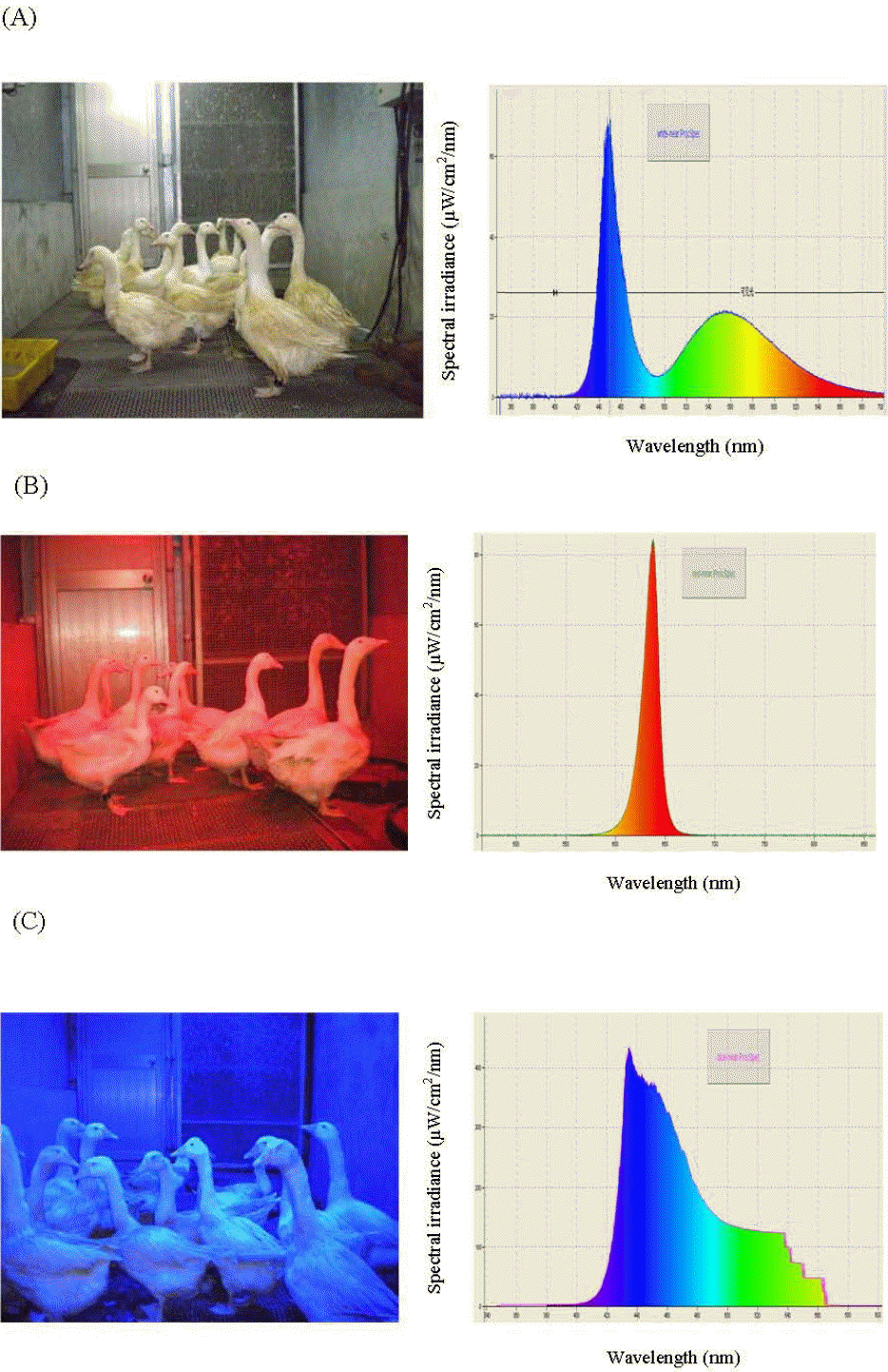
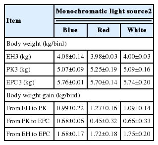
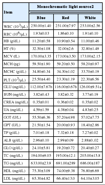
The monochromatic light and wavelength in goose house. (A) Geese rearing under white light-emitting diode (LED)-light and spectral sensitivity; (B) Geese rearing under red LED-light and spectral sensitivity; (C) Geese rearing under blue LED-light and spectral sensitivity.
Growth performance
The body weight and feed consumption were measured and recorded immediately after the birds entered the environmentally controlled house. The peak of egg production was defined as the average of laying rate for 2 consecutive days reached 35% for all the geese. The peak reaching at approximately 8 weeks after geese entered the environment controlled house. The egg production ceased (EPC) at approximately 38 weeks after geese entered the house. The EPC was defined as the average of laying rate for 2 consecutive days dropping to 5% for all the geese.
Fertilization rate and hatchability
A total of 13,077 eggs were collected from the 3 blocks. To determine fertilization rate, hatchability and gosling output, eggs were collected in batches every 2 weeks from July 26, 2011 to January 20, 2014. Eggs were incubated with an automatic incubator (Tonz Nan Incubators Manufactured Co. Ltd., Yunlin, Taiwan). The temperature of the incubator was set at 37.7°C from day 0 to 14, 37.5°C from days 15 to 28, and 37.2°C during hatching. The transparency of the fertilized eggs was judged with the naked eye on the 7th d of incubation. The percentage of fertilized eggs served as the fertilization rate. Hatchability was defined as the percentage of goslings hatched over the number of total eggs incubated. The hatchability of fertilized eggs was defined as the percentage of goslings hatched over the total number of fertilized eggs.
Egg measurements
Eggs were transferred to the laboratory and stored in a refrigerator until analysis (within two days of collection). Egg weight, eggshell shape index (ESI), egg length, egg width, eggshell strength, and eggshell thickness were recorded once every month. Egg weight was measured to the nearest 0.01 g using an electronic balance. Egg length and egg width at midpoint on the outer surface of the egg were measured in micrometers (0.01 mm, Mitutoyo, Kawasaki, Japan), and ESI was calculated using the formula ESI = width/length×100, where width is the transverse diameter, and length is the long vertical length of an egg.
Serum biochemical parameters
During the experiment periods, 4 geese (2 male and 2 female 10-month-old geese) per pen were randomly chosen for collecting serum samples for biochemical determinations at the first day at each month. Blood samples were processed approximately 4 to 5 h after collection by centrifuging at 3,000×g for 10 min at 4°C, and serum samples were stored at −4°C for up to 1 d until analysis (Lee et al., 2013). Serum biochemical parameters analyses were performed by an Automatic Biochemical Analyzer (Hitachi, 7150 auto-analyzer, Tokyo, Japan). For reproductive hormones assay, the serum was analyzed by electrochemiluminescence immunoassay (ECLIA) using an Elecsys and cobase immunoassay analyzer (UniCel DxI 800 Access Immunoassay System, Beckman Coulter Inc., Fullerton, CA, USA).
Measurement of serum melatonin concentration
Blood samples collected from geese that had adapted to the dark for 6 h were then centrifuged at 3,000×g for 15 min. The serum melatonin concentration was measured using a commercial enzyme-linked immunosorbent assay (ELISA) kit (USCN Life Science Inc., Wuhan, China) as per the manufacturer’s protocol. Standard curve and regression equations were used; based on the degree of fitness, as well as the conciseness of the regression curve, a quadratic equation was ultimately selected. Using this equation, the melatonin concentration of all of the samples was calculated with an ELISA reader (Sunrise-Basic-Tecan, Grödig, Austria) set to an optical density at 450 nm and a standard range of 4.940 to 10,000 pg/L.
Statistical analysis
The data were statistically analyzed using the general linear model procedure (GLM) of SAS software (2004) following a random arrangement. The mathematical model was:
where, Yijk = observed response of bird in a pen; Ti = fixed effect of colored light treatment; Bj = fixed effect of batch; ɛijk = residual error when the pen was regarded as an experimental unit,
RESULTS
Growth performance and serum biochemical parameters
The effect of monochromatic light on the growth of breeder geese is shown in Table 2. There was no significant difference in the body weight among the three lighting groups throughout the experimental period, from the point in time when the birds entered the house, through peak egg production, until egg production ceased (p>0.05). The effect of monochromatic light on blood biochemical parameters in breeder geese is presented in Table 3. The glucose level in the white light group was significantly higher than that in the red and blue light groups (p<0.05). Other blood biochemical parameters did not differ among the treatment groups (p>0.05).
Reproductive performance
The effect of monochromatic light on the reproductive characteristics of breeder geese is shown in Table 4. The eggs in the red and white light groups were significantly more numerous than those of the blue light group (p<0.05). The laying period for the red light group was also significantly longer than that of the white and blue light groups (p<0.05).
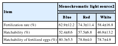
Fertilization rate and hatchability
The effect of monochromatic light on the fertilization rate and hatchability of breeder geese is shown in Table 5. The fertilization rate, the hatchability and the hatchability of fertilized eggs in the blue light group was similar to those of the other two groups (p>0.05).
Egg measurements
The effect of monochromatic light on egg measurements in breeder geese is shown in Table 6. The lengths of the eggs in the red and white light groups were significantly longer than those of the blue light group (p<0.05). Accordingly, the egg shape indices for the red and white light groups were significantly lower than that of the blue light group (p<0.05).

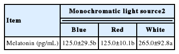
Serum melatonin concentration
The effect of monochromatic light on the concentration of melatonin in the serum of the breeder geese is shown in Table 7. Melatonin levels were significantly higher in the white light group compared to those of the blue and red light groups (p<0.05).
DISCUSSION
The eyes and the extra-retinal photoreceptors located in the hypothalamus or other sites of the brain are sensitive to different light wavelengths, and are involved in photostimulation. Therefore, we designed 30 lux light intensity in this study during the experiment. No obvious difference on the performance was observed.
Rozenboim et al. (1999) reported that broilers reared under blue or green light were significantly heavier than those reared under white or red light. This suggests that blue or green light may have an effect on the number of satellite cells per gram of breast muscle, as well as the total number of satellite cells derived from the broilers (Halevy et al., 1998). In contrast, the results in this study showed that there was no significant difference in either body weight or body weight gain among different lighting groups during the laying period. Moreover, there were also no significant differences in the laying rate and survival rate among birds in the blue, red, and white light groups during the laying period. However, Rozenboim et al. (1999) reported that blue light enhancement of growth was probably due to an elevation in plasma androgens at later age in the broilers. Therefore, the authors suggested that lower light intensity, as well as specific light spectra, could enhance the growth of muscles. The discrepancy between our results and those of Rozenboim et al. (1999) may be caused by differences in species, age, and experimental conditions applied.
Hassan et al. (2013) found that laying hens under red light had higher egg numbers compared to those under blue or green light. The authors suggested that hens in the red light group had significantly higher organ weights (stroma of ovaries) and ovarian follicle numbers (1 to 3 and 4 to 6 mm). In the present study, the red light group also had a significantly higher egg number than that of the blue light group, that is to say the red light group had a longer laying period than the blue or white light groups, suggesting a prolonged period of onset to laying peak. Serum glucose level is a stress indicator and birds under stress condition have elevated the serum parameter (Puvadolpirod and Thaxton, 2000). Hassan et al. (2013) showed that hens under red light had significantly higher serum glucose and triglyceride levels than those under white or blue light. However, Firouzi et al. (2014) showed that birds under red and blue lights had lower levels of serum glucose than yellow and green lights and the authors also demonstrated that wavelength had calming effect on broilers (Prayitno et al., 1997). The observed is in agreement with our results.
Monochromatic LED light colors were tested and it was found that red light enhances egg production of laying hens (Kim et al., 2012; Hassan et al., 2014). In this study, we found that the breeder geese under red light showed a higher fertilization rate than that of the white light group, the result is in agreement with Li et al. (2014). The results suggested that the birds under red light had increased follicle-stimulating hormone and estradiol (E2) concentrations compared to the blue light group (Hassan et al., 2013). Furthermore, Gongruttananun and Guntapa (2012) also showed that hens under red light had a significantly higher concentration of serum estradiol compared to the hens exposed to daylight supplemented with fluorescent light.
Laying hens under red light had lower average egg weight during the laying period; the average egg length was also lower than those under blue, green or white light (Er et al., 2007). However, Gongruttananun and Guntapa (2012) found no significant difference in average egg weight, egg or eggshell quality between hens kept under red light or those exposed to natural daylight supplemented with fluorescent light. The present study did not find any difference in average egg weight or egg width, but the blue light group had a significantly greater egg shape index compared to those in the other groups. This result depended on egg length in blue light becoming shorter than in red and white lights, and the result is in agreement with Li et al. (2014) who reported that the egg length in blue light was significantly shorter than red and white lights, but no difference in egg width in laying hens.
Melatonin is synthesized from serotonin; it can stimulate gonadotropin-inhibitory hormone (GnIH) secretion which may ultimately inhibit reproduction (Ubuka et al., 2005). Due to its effect on the anterior pituitary gland and the gonadotropin-releasing hormone system, GnIH can regulate avian reproduction by inhibiting gonadotropin release and synthesis (Bentley et al., 2006). Jin et al. (2011) indicated that the concentration of plasma melatonin under green light was the highest, followed by white light, and was the lowest in red light and blue light for broilers. Gordijn et al. (2005) showed that the melatonin increase in the evening was significantly suppressed under blue light as compared to the red light. In fact, the concentration of serum melatonin under red light is 3.8-fold higher than the melatonin under blue light. Huang (2008) also concluded that long photoperiod causes earlier cessation of laying and the effect cannot be abolished by melatonin feeding. It is implied that melatonin is not the main mediator for the effect of photoperiod on reproduction in geese. Therefore, we suggest that melatonin is not the main mediator of reproduction in breeding geese.
CONCLUSION
Our results suggested that red LED-light has the best effect on reproductive performance (i.e. longer laying period and higher total eggs number) at 30 lux light intensity, and is therefore a better choice for the management of breeding geese than blue or white LED-light.
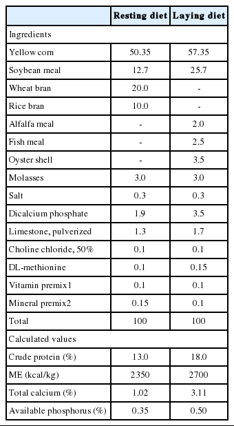
Ingredients and nutrient compositions of the experimental diets (as fed basis)
ACKNOWLEDGMENTS
The authors would like to thank colleagues in the Changhua Animal Propagation Station, COA-LRI in Taiwan, for managing the breeder geese.
Notes
CONFLICT OF INTEREST
We certify that there is no conflict of interest with any financial organization regarding the material discussed in the manuscript.