


INTRODUCTION
The assessment of milk microbiota is important for preventing mastitis and maintaining herd health [1ŌĆō3]. If typical contagious bacteria, such as Staphylococcus aureus (S. aureus), Streptococcus agalactiae (S. agalactiae), and Corynebacterium bovis (C. bovis), are found in the tank milk, the infected cows should be identified, and the procedure and sequence of milking should be revised. If environmental bacteria, such as coliforms, coagulase-negative Staphylococci, and Streptococci other than S. agalactiae, markedly increase, the hygiene of the cowshed should be improved. Likewise, pathogen identification greatly facilitates the provision of appropriate treatment with antibiotics. Regarding the quality control of milk, the abundance of Pseudomonas spp. may be of great concern, because their heat-resistant enzymes can hydrolyze protein and fat, producing an unpleasant flavor even after pasteurization [4,5]. Moreover, several Pseudomonas spp. cause mastitis [6].
The prevalence of mastitis varies according to the season in dairy cows as does the somatic cell count (SCC), which is an indicator of the number of leukocytes in the milk and therefore of udder health. Makovec and Ruegg [7] reported that mastitis caused by Escherichia coli (E. coli) and Streptococcus spp. may occur more during summer, and Olde Riekerink et al [2] described the greater risk of mastitis caused by S. aureus and Streptococcus dysgalactiae during winter. Olde Riekerink et al [2] also reported the effect of housing on seasonal variation; mastitis caused by E. coli can occur more during summer if cows are managed on pasture, whereas the infection may be seen more during winter if managed as confined herds. Mastitis caused by coagulase-negative S. aureus and C. bovis may occur throughout the year. Milk spoilage caused by psychrophilic Pseudomonas spp. can become a problem during winter.
Regardless of the symptoms of mastitis, the milk microbiota varies between seasons [8]. Differences in temperature and humidity could account for the variation, because both the health of the cows and the growth of milk bacteria are influenced by temperature and humidity. However, Li et al [9] found that at the phylum level the milk microbiota during summer was similar to that during winter. In fact, it is unclear what causes seasonal variations in the milk microbiota.
Among the factors putatively involved in the milk microbiota and mastitis outbreak, the bedding microbiota may be the most important because the teats of a dairy cow are in direct contact with bedding material when the cow is resting. In a previous study, we examined the microbiota of the gut, milk, and cowshed environment using 16S rRNA gene amplicon sequencing, and demonstrated that the milk microbiota is associated with the bedding microbiota but is clearly distinct from the feed, rumen fluid, fecal, and water microbiota [10]. We conducted the survey at two farms: one in April and one in September; hence, the difference in the milk microbiota between the farms might have resulted from seasonal variations. Although the importance of the bedding microbiota has long been recognized from the perspective of mastitis prevention, few studies have examined the milk and cowshed microbiota or the variation across seasons.
In the present study, we collected samples of milk, feces, bedding, and airborne dust from a dairy farm during summer and winter at 1 and 2 months postpartum. The microbiota was assessed by 16S rRNA gene amplicon sequencing. The objective was to determine if the milk and cowshed microbiota vary between seasons, and if a variation in the milk microbiota is related to a variation in the cowshed microbiota.
MATERIALS AND METHODS
Sampling
We collected samples from cows at the Okayama Prefecture Livestock Research Institute (Okayama, Japan). The cows were housed in a free stall barn and fed a total mixed ration silage throughout the year (Table 1). During the summer their diet was supplemented with a fatty acid salt (palmitic acid calcium) to fortify milk fat production. The contents of dry matter (DM), crude protein (N├Ś6.25), and total digestible nutrients were 55% to 60%, 16% to 17%, and 72% to 4% DM, respectively. The sampling was performed from 6 June to 22 August and from 17 November to 16 January. Hereafter, the former series is referred to as the summer sampling and the latter as the winter sampling. The minimum and maximum temperatures were 16┬░C to 31┬░C during the summer and ŌłÆ2┬░C to 15┬░C during the winter.
The milk and feces samples were collected from 9 cows during summer and from 8 cows during winter, with 2 sampling times each at 1 and 2 months postpartum. Because milk and fecal sampling from cows at 1 and 2 months postpartum was occasionally conducted on the same day, bedding and airborne dust sampling was carried out 6 times during summer and 9 times during winter.
Each milk sample was collected after cleaning the surface of the udder, and the foremilk was discarded before collecting the sample. Milk samples were taken manually from 4 udders, then mixed to produce a composite sample. Fecal samples were collected from the rectum. Airborne dust samples were collected by placing 3 petri dishes approximately 1.0 m above the ground for 5 min. Bedding samples were collected from 3 separate places in a cowshed. In the free stall system, cows can move and rest freely, and determining their resting place was difficult. Thus, a composite sample prepared from 3 separate samples was regarded as a representative means of assessing the bedding and airborne dust microbiota at the time of sampling. The institute operated automatic milking systems (Lely Astronout A4, Cornes AG. Ltd., Eniwa, Japan) that enabled cows to be milked at any time; hence, although all samplings were completed between 10:00 and 12:00, the time when we collected the milk after milking was variable. All the samples were kept on ice during transportation to the laboratory and were stored at ŌłÆ20┬░C until required for further analyses. Overall, 34 milk, 34 feces, 15 bedding, and 15 airborne dust samples were subjected to MiSeq analysis.
The contents of protein, fat, and solids-not-fat (SNF), and the SCC of the milk were determined using a CombiFoss FT+ analyzer (Foss All├®, Hiller├Ėd, Denmark). The procedures and protocols for the animal experiments were approved by the Animal Care and Use Committee, Okayama University (OKU-2016290), Japan.
DNA extraction
The 250 ╬╝L milk samples were centrifuged at 16,000 g for 2 min, and the pellet was collected. For DNA extraction from airborne samples, a 1-mL aliquot of each sample was transferred into an Eppendorf tube, and then centrifuged to collect the pellet. All the pellet samples were washed with 500 ╬╝L of solution I containing 0.05 M D-glucose, 0.025 M Tris-HCl (pH 8.0), and 0.01 M sodium ethylenediaminetetraacetic acid (EDTA) (pH 8.0), and then lysed with 180 ╬╝L of lysozyme solution (20 g/L lysozyme, 0.02 M Tris-HCl [pH 8.0], 0.002 M sodium EDTA [pH 8.0], 1.2 g/L Triton X-100) at 37┬░C for 1 h. Bacterial DNA of milk, airborne dust samples was purified by using the DNeasy blood & tissue kit (Qiagen, Germantown, MD, USA), according to the manufacturerŌĆÖs instructions. For feces and bedding samples, a 0.2 g of the sample was used for bacterial DNA extraction following the procedure for the repeated bead beating plus column method [11] and purified using the mini DNeasy stool kit (Qiagen, USA).
16S rRNA gene amplicon sequencing
Bacterial DNA was amplified by two-step polymerase chain reaction (PCR) to generate amplicon libraries for next-generation sequencing [12]. The primers targeting the V4 region of 16S ribosomal RNA (rRNA) genes (forward: 5ŌĆ▓-ACACTC TTTCCCTACACGACGCTCTTCCGATCTGTGCCAGCMGCCGCGGTAA-3ŌĆ▓; reverse: 5ŌĆ▓-GTGACTGGAGTTCA GACGTGTGCTCTTCCGATCTGGACTACHVGGGTW TCTAAT-3ŌĆ▓) were used for the first round of PCR, with the following protocol: initial denaturation at 94┬░C for 2 min, followed by 35 cycles of denaturation at 94┬░C for 30 s, annealing at 50┬░C for 30 s, elongation at 72┬░C for 30 s, and an elongation step at 72┬░C for five min. The PCR products were purified by electrophoretic separation on a 2.0% agarose gel using a Fast Gene Gel/PCR Extraction Kit (NIPPON Genetics Co., LTD., Tokyo, Japan). The second round of PCR, with adapter-attached primers, followed the protocol of initial denaturation at 94┬░C for two min, 10 cycles of denaturation at 94┬░C for 30 s, annealing at 59┬░C for 30 s, elongation at 72┬░C for 30 s, and an elongation step at 72┬░C for five min. The second-round PCR products were purified in the same way as that in case of the first-round PCR products.
The purified amplicons were pair-end sequenced (2├Ś250 bp) on an Illumina MiSeq platform at FASMAC Co., Ltd. (Kanagawa, Japan). Raw sequence data were analyzed using the Quantitative Insights Into Microbial Ecology (QIIME version 1.9.0). The 250-bp reads were truncated at any site receiving an average quality score under 20. Truncated reads that were shorter than 225 bp were discarded. In primer matching, sequences showing overlaps longer than 200 bp were assembled. The final reads obtained after pair-end joining were grouped into operational taxonomic units using a 97% similarity threshold. The sequence data were analyzed and categorized from the phylum to the family level using the default settings of the Ribosomal Database Project classifier.
Statistical analysis
Data pertaining to the milk yields, milk composition, blood metabolite concentrations, and relative abundances of major bacterial families in the milk, feces, bedding, and airborne dust microbiota (where the proportion of the family in at least one sample was >1.0%) were analyzed by the non-parametric MannŌĆōWhitney U test to examine the effects of seasons and months after calving. The microbiota data were also subjected to canonical analysis of principal coordinates (CAP) to define assignment and clustering that explained the variations in the microbiota. Discriminant vectors with a Pearson correlation >0.7 were considered significant. The non-parametric test was performed using JMP software (version 11; SAS Institute, Tokyo, Japan) and CAP was carried out using Primer version 7 with the Permanova+ add-on (Primer-E, Plymouth Marine Laboratory, Plymouth, UK).
RESULTS
The milk yield of the cows was 35 to 39 kg, and there were no differences between the two seasons or between 1 and 2 months after calving (Table 2). The content of milk protein was greater, and the contents of fat and SNF were numerically higher during winter than during summer. The average SCC of the milk at 2 months postpartum during summer reached 429├Ś103 cells/mL, because one cow had a markedly high SCC (2.8├Ś106 cells/mL).
At the family level, the five most abundant taxa of the milk microbiota during summer were Staphylococcaceae (10.3%), Ruminococcaceae (9.7%), Aerococcaceae (7.7%), Lachnospiraceae (5.4%), and Corynebacteriaceae (4.3%), and those during winter were Ruminococcaceae (10.2%), Staphylococcaceae (7.5%), Lactobacillaceae (6.5%), Aerococcaceae (5.5%), and Lachnospiraceae (5.3%) (Table 1). There were seasonal variations in the relative abundances of Bacillaceae, Micrococcaceae, and Staphylococcaceae (summer > winter) and those of Streptococcaceae, and Microbacteriaceae (summer < winter). There were also variations in the relative abundances of Porphyromonadaceae (1 month > 2 months), and Turicibacteraceae and Tissierellaceae (1 month < 2 months) between samples taken 1 or 2 months postpartum.
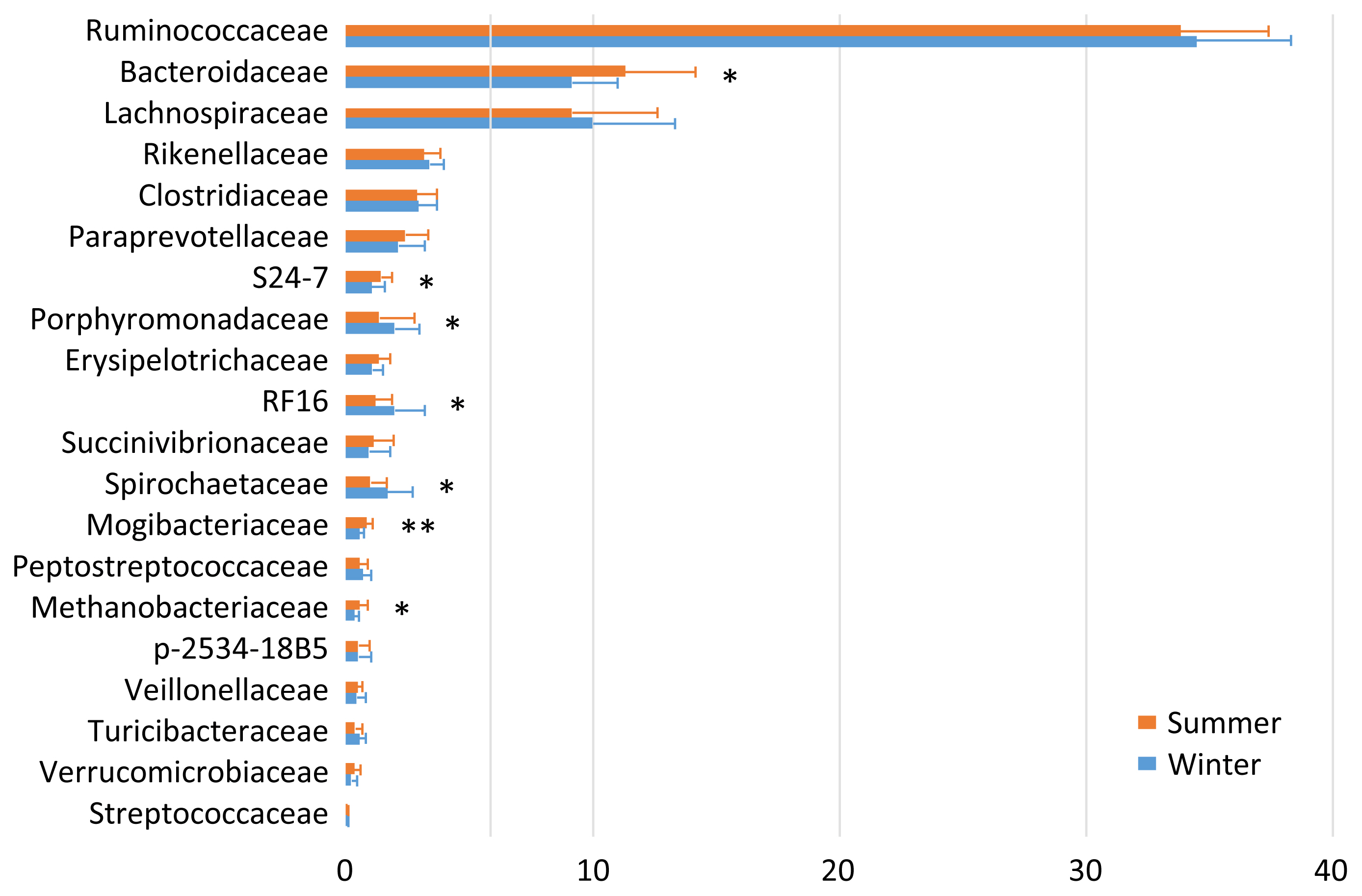
The fecal microbiota was fairly stable at 1 and 2 months postpartum. Therefore, the data are summed up to enable comparison between the two seasons (Figure 1). Ruminococcaceae (34.1%), Bacteroidaceae (10.2%), Lachnospiraceae (9.6%), Rikenellaceae (3.3%), and Clostridiaceae (2.9%) were the five most abundant taxa regardless of the season. There were seasonal variations in the relative abundances of S24-7, Mogibacteriaceae, and Methanobacteriaceae (summer > winter), and of Porphyromonadaceae, RF16, and Spirochaetaceae (summer < winter).
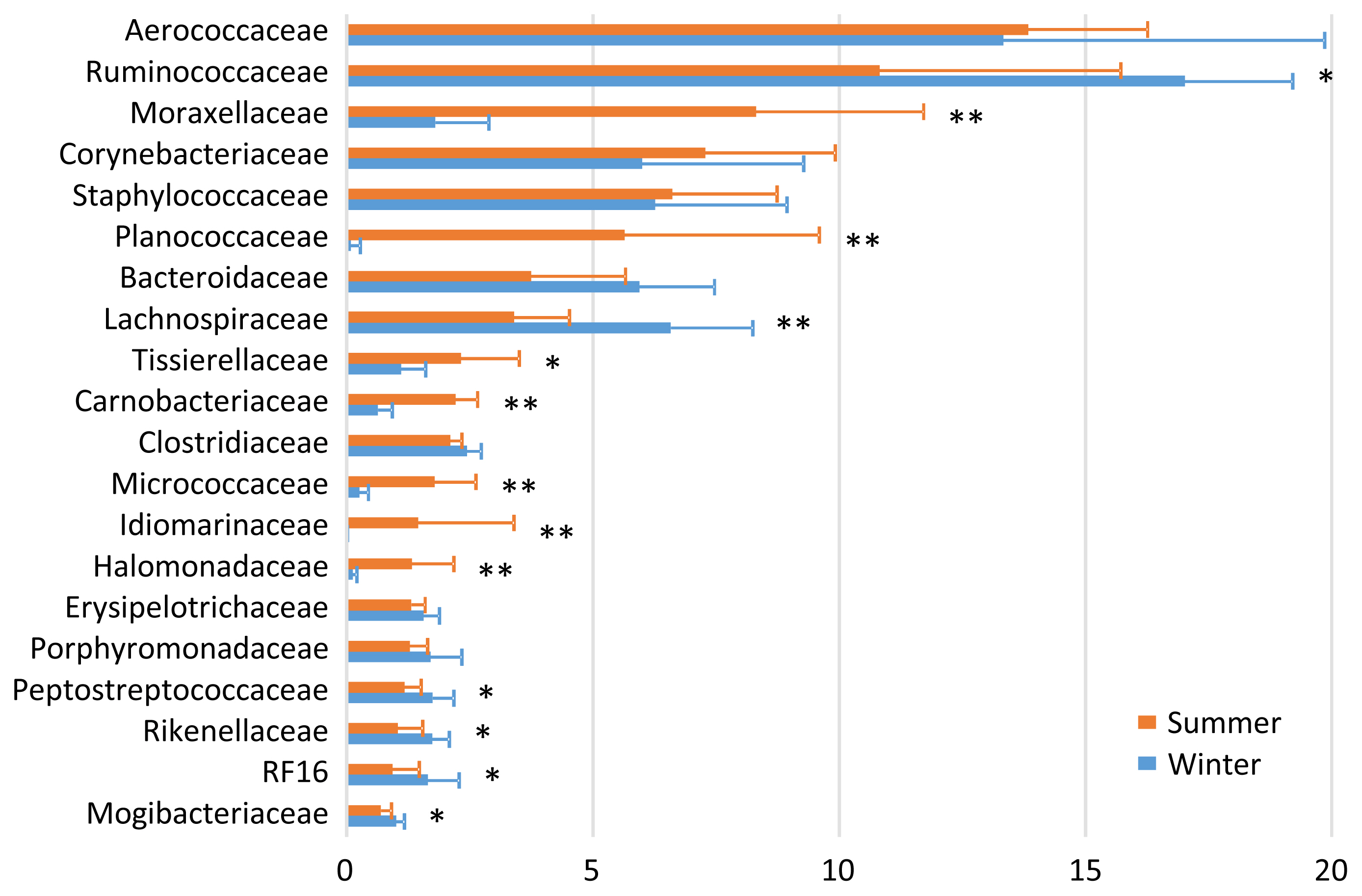
In the bedding microbiota, Aerococcaceae (13.8%), Ruminococcaceae (10.8%), Moraxellaceae (8.3%), Corynebacteriaceae (7.3%), and Staphylococcaceae (6.6%) were the five most abundant taxa during summer, and Ruminococcaceae (17.0%), Aerococcaceae (13.3%), Lachnospiraceae (6.6%), Staphylococcaceae (6.3%), and Corynebacteriaceae (6.0%) were the five most abundance taxa during winter (Figure 2). There were seasonal variations in the relative abundances of Moraxellaceae, Planococcaceae, Tissierellaceae, Carnobacteriaceae, Micrococcaceae, Idiomarinaceae, and Halomonadaceae (summer > winter); and of Ruminococcaceae, Lachnospiraceae, Peptostreptococcaceae, Rikenellaceae, RF16, and Mogibacteriaceae (summer < winter).
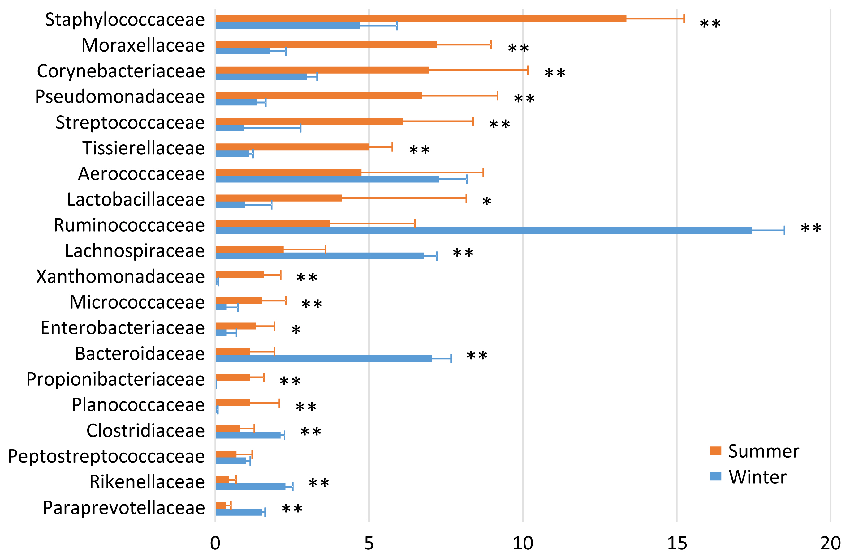
In the airborne dust microbiota, the five most abundant taxa during summer were Staphylococcaceae (13.4%), Moraxellaceae (7.2%), Corynebacteriaceae (7.0%), Pseudomonadaceae (6.7%), and Streptococcaceae (6.1%); and the five most abundant taxa during winter were Ruminococcaceae (17.4%), Aerococcaceae (7.3%), Bacteroidaceae (7.1%), Lachnospiraceae (6.8%), and Staphylococcaceae (4.7%) (Figure 3). There were seasonal variations in the relative abundances of Staphylococcaceae, Moraxellaceae, Corynebacteriaceae, Pseudomonadaceae, Streptococcaceae, Tissierellaceae, Lactobacillaceae, Xanthomonadaceae, Micrococcacae, Enterobacteriaceae, Propionibacteriaceae, and Planococcaceae (summer > winter); and of Ruminococcaceae, Lachnospiraceae, Bacteroidaceae, Clostridiaceae, Rikenellaceae, and Paraprevotellaceae (summer < winter).
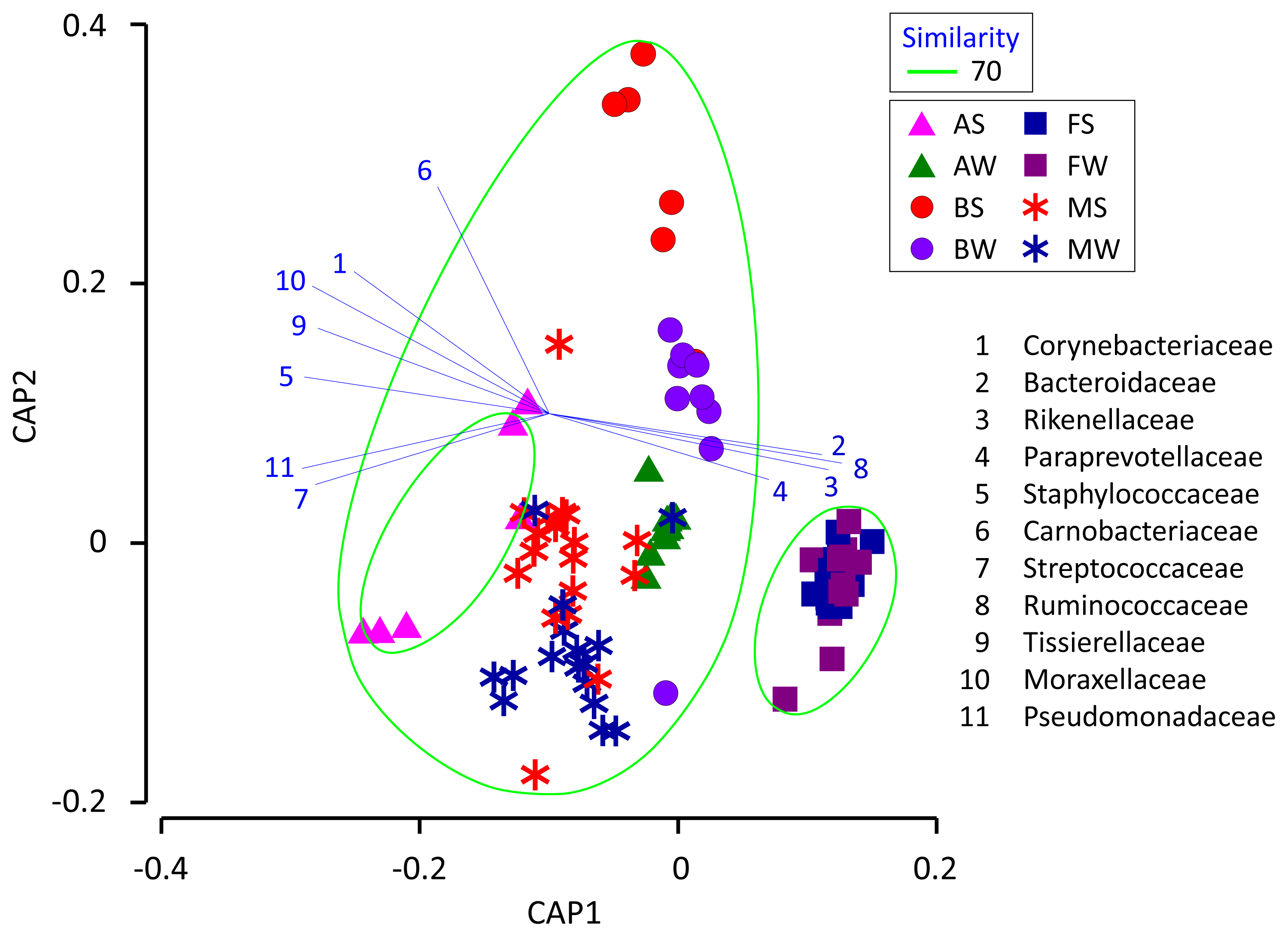
We used CAP to determine if the milk microbiota was related to the cowshed microbiota (Figure 4). The results indicate that the milk microbiota was grouped together with the bedding and airborne dust microbiota, and not with the fecal microbiota. Likewise, several airborne dust samples taken during summer were low in Ruminococcaceae, Bacteroidaceae, and Lachnospiraceae, and formed a separate group from the others.
DISCUSSION
Milk yield and protein content follow a seasonal pattern over the course of the year [13]. They are typically greatest during winter and reach a nadir during summer; thus, the higher protein content of the milk obtained during winter was regarded as normal. The dairy cows investigated in the present study were given the same diet (total mixed ration silage) throughout the year, except during the summer when their diet was supplemented with a small amount of palmitic acid calcium. It is therefore difficult to explain why the relative abundances of S24-7, Mogibacteriaceae, and Methanobacteriaceae were greater, and those of Porphyromonadaceae, RF16, and Spirochaetaceae were lower during summer in the fecal microbiota.
Regardless of whether the cows had normal or high SCC, Metzger et al [8] found variation of the milk microbiota between the sampling times after calving, i.e., 1 week, 2 weeks, and 2, 3, 4, or 5 months of lactation. In the present study, only Porphyromonadaceae (decrease), Turicibacteraceae (increase), and Tissierellaceae (increase) exhibited changes in abundance at 1 to 2 months postpartum. This differed from the finding of Metzger et al [8], wherein the abundancies of numerous bacterial taxa varied between sampling times.
Metzger et al [8] also demonstrated that variation of the milk microbiota was greater between seasons than between the sampling times after calving; the abundancies of a large number of bacterial taxa including Staphylococcus spp., Acinetobacter spp., and Aerococcus spp. varied between the seasons. In the present study, only five families exhibited seasonal variation; the abundances of Micrococcaceae, Bacillaceae, and Staphylococcaceae were greater during summer, and Microbacteriaceae and Streptococcaceae were more prevalent during winter. Li et al [9] reported that the abundances of Pseudomonas spp., Propionibacterium spp., and Flavobacterium spp. were negatively correlated with temperature, and those of Bacillus spp., Lactobacillus spp., and Bifidobacterium spp. were positively correlated with temperature. In the present study, we observed a similar temperature effect to that described by Li et al [9] for Bacillaceae (summer > winter).
Regarding the relationships between the milk and cowshed microbiota, the pattern of seasonal variation was the same between the milk and bedding samples for Micrococcaceae (summer > winter), and between the milk and airborne dust samples for Staphylococcaceae and Micrococcaceae (summer > winter). The pattern was reversed between the milk and airborne dust samples for Streptococcaceae. Regarding the relationships between the milk and fecal microbiota, the patterns of seasonal variation (summer > winter or summer < winter) was not the same for any families.
The finding that the milk microbiota was related to the bedding and airborne dust microbiota agreed with the results reported by Wu et al [10]. Although the typical bacterial taxa of feces, i.e., Ruminococcaceae, Bacteroidaceae, Lachnospiraceae, Clostridiaceae, and Rikenellaceae, were stably detected in the milk microbiota, Aerococcaceae, Staphylococcaceae, Moraxellaceae, Corynebacteriaceae, Streptococcaceae, Pseudomonadaceae, and Tissierellaceae, which are regarded as typical bacterial taxa of bedding and airborne dust, were not abundant in the fecal microbiota. Interestingly, Lachnospiraceae and Aerococcaceae were not included in the discriminant vectors with a Pearson correlation >0.7 in the CAP. Instead, Carnobacteriaceae contributed to the differentiated groups. Although the relative abundance of Carnobacteriaceae was <1.0% in all the milk samples, the pattern of seasonal variation (summer > winter) was the same in the milk and bedding samples. Carnobacteriaceae is known as a spoilage-associated taxon in meat [14], although its significance regarding milk quality has yet to be elucidated.
Even though the seasonal variation of the bedding and airborne dust microbiota did not influence the milk microbiota, a distinctive group comprising the milk, bedding, and airborne dust microbiota was formed by the CAP. Therefore, seasonal variation of the milk microbiota mat result from factors that have greater influences than the seasonal variation of the cowshed microbiota. Further research is required to clarify the factors involved in the seasonal variation of the milk microbiota.
Although the importance of the cowshed microbiota has long been recognized, few researchers have investigated the airborne dust microbiota of a dairy farm. Dutkiewicz et al [15] examined cowshed microbiota by plate culture, and isolated numerous species including those belonging to the following genera: Micrococcus, Arthrobacter, Staphylococcus, Bacillus, Corynebacterium, Microbacterium, Streptomyces, Acinetobacter, Proteus, Pantoea, Pseudomonas, Thermoactinomyces, and Saccharopolyspora. Although most of the corresponding families, i.e., Micrococcaceae, Staphylococcaceae, Bacillaceae, Corynebacteriaceae, Microbacteriaceae, Moraxellaceae, Enterobacteriaceae, and Pseudomonadaceae, were detected by amplicon sequencing, the abundance of Streptomycetaceae was quite low (<0.01%), and we did not detect Thermoactinomycetaceae in the present study.
The distinctive seasonal variation in the bedding and airborne dust microbiota is difficult to explain, because few relevant surveys have been performed. The dairy farm used fans with a mist of water to cool the bodies of the cows during summer; hence, this enforced ventilation may have caused the differences in the bedding and airborne dust microbiota between the two seasons. The lower abundances during summer than during winter of the five typical bacterial taxa of feces, i.e., Ruminococcaceae, Bacteroidaceae, Lachnospiraceae, Clostridiaceae, and Rikenellaceae, may have resulted from the water mist ventilation.
 PDF Links
 PubReader
 ePub Link
 Full text via DOI
 Download Citation
 Print
Print


